. 2022 Jan;50(1):1-15.
doi: 10.1007/s10439-021-02882-0. Epub 2022 Jan 7.

Patient-Specific Quantification of Normal and Bicuspid Aortic Valve Leaflet Deformations from Clinically Derived Images

Affiliations

Patient-Specific Quantification of Normal and Bicuspid Aortic Valve Leaflet Deformations from Clinically Derived Images

Bruno V Rego et al. Ann Biomed Eng. 2022 Jan.

Abstract

The clinical benefit of patient-specific modeling of heart valve disease remains an unrealized goal, often a result of our limited understanding of the in vivo milieu. This is particularly true in assessing bicuspid aortic valve (BAV) disease, the most common cardiac congenital defect in humans, which leads to premature and severe aortic stenosis or insufficiency (AS/AI). However, assessment of BAV risk for AS/AI on a patient-specific basis is hampered by the substantial degree of anatomic and functional variations that remain largely unknown. The present study was undertaken to utilize a noninvasive computational pipeline ( https://doi.org/10.1002/cnm.3142 ) that directly yields local heart valve leaflet deformation information using patient-specific real-time three-dimensional echocardiographic imaging (rt-3DE) data. Imaging data was collected for patients with normal tricuspid aortic valve (TAV, [Formula: see text]) and those with BAV ([Formula: see text] with fused left and right coronary leaflets and [Formula: see text] with fused right and non-coronary leaflets), from which the medial surface of each leaflet was extracted. The resulting deformation analysis resulted in, for the first time, quantified differences between the in vivo functional deformations of the TAV and BAV leaflets. Our approach was able to capture the complex, heterogeneous surface deformation fields in both TAV and BAV leaflets. We were able to identify and quantify differences in stretch patterns between leaflet types, and found in particular that stretches experienced by BAV leaflets during closure differ from those of TAV leaflets in terms of both heterogeneity as well as overall magnitude. Deformation is a key parameter in the clinical assessment of valvular function, and serves as a direct means to determine regional variations in structure and function. This study is an essential step toward patient-specific assessment of BAV based on correlating leaflet deformation and AS/AI progression, as it provides a means for assessing patient-specific stretch patterns.

Keywords: Aortic valve; Computational modeling; Deformation; Echocardiography; Patient stratification.


Figures

FIGURE 9.
FIGURE 9.
In-plane stretch in the normal TAV, showing the capability of the method to capture substantial regional heterogeneity as well as directional differences in stretch.
FIGURE 1.
FIGURE 1.
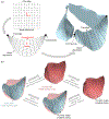
Pipeline for AV geometric modeling on a patient-specific basis, starting from noninvasive rt-3DE images of the valve. Full target geometries of the AV obtained through image segmentation (a) are processed using an established deformable medial modeling approach (b), which yields leaflet-specific triangulated mesh models of the leaflet medial surface (c). In addition to describing the AV leaflet medial surface geometry, each leaflet’s geometric model is enriched with a local radius function (d), which defines the pointwise thickness of the leaflet.
FIGURE 2.
FIGURE 2.
Detailed methodology for converting segmented rt-3DE images to a cm-rep of each leaflet. (a) Segmentations of the leaflets in the rt-3DE image were obtained. (b) The raw rt-3DE segmentation of each leaflet was smoothed with a Gaussian kernel. (c) 3D Voronoi skeletonization was performed to obtain an approximate medial surface of the segmented leaflet and a triangulated mesh of the medial surface was manually generated on the Voronoi skeleton. This mesh served as an initialization, or deformable template, for medial modeling of the leaflet. We defined all AV leaflets in this study to have a non-branching medial axis. In fused BAV leaflets, we manually labeled the mesh triangles located at the raphe. (d) To obtain the final medial axis representation, the template was refined using Loop subdivision. (e) The coordinates and associated radius (r) value at each node were optimized to maximize the volumetric overlap between the smoothed segmentation and a closed boundary reconstructed from the medial model as described in Ref. [20].
FIGURE 3.
FIGURE 3.
Pipeline for acquiring the deformed (i.e. closed, fully loaded) state on a per-leaflet basis. (a) First, the open- and closed-state medial surface geometries are acquired from rt-3DE (Figs. 1 and 2), shown here for a TAV. (b) Throughout the FE simulation, boundary displacement, bulk leaflet pressurization, and shape enforcement are precisely coordinated to ensure accuracy and stability. (c) During closure, the loading conditions on the leaflet are adjusted locally using a corrective pressure field that is proportional to the signed distance d between any point and the nearest location on the true closed surface. This local pressure continually pushes the simulated leaflet surface toward the true closed configuration and thus enforces the true closed shape by the end of the simulation. Representative surface geometries and cross sections are shown for the open state, the onset of shape enforcement, the final simulated closed state, and the true closed state.
FIGURE 4.
FIGURE 4.
(a) Spline surfaces are defined parametrically in R2 based on the spline coordinates (u, v), from which circumferential and radial directions are defined locally. This spline surface can then be morphed to the leaflet surface in 3D via least-squares fitting, to allow projection of stretch results onto the anatomic directions and averaging of results across subjects for each leaflet type. (b) Pipeline for enforcing material point correspondence between open- and closed-state spline surfaces. Nodes on the open and closed triangulated leaflet meshes are treated as material points (i.e. correspondent between configurations), due to the fact that the closed-state nodes are obtained through a FE simulation, which uses finite elasticity to determine the deformed nodal positions. To maintain this correspondence between the open and closed spline surfaces, the parametric locations of the mesh nodes in the open state were maintained using equality constraints during the closed-state fitting process.
FIGURE 5.
FIGURE 5.
Representative stretch fields at full closure for a TAV and a BAV, showing local maximum and minimum in-plane stretch magnitudes and directions. Differences by leaflet type are qualitatively apparent; note especially that in the raphe of the BAV, the maximum in-plane stretch is substantially lower than over the rest of the fused leaflet.
FIGURE 6.
FIGURE 6.
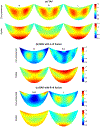
Key study results, showing mean circumferential and radial stretch fields for (a) TAV patients, (b) BAV patients with L–R fusion, and (c) BAV patients with R–N fusion, averaged across each leaflet type using the spline surface parameterization. Note the large differences between the BAV leaflets and their TAV counterparts, as well as the visible effect of the raphe (central) region on the circumferential stretch field of L–R (R–L) and R–N (N–R) leaflets.
FIGURE 7.
FIGURE 7.
Group-wise stretches in (a) the principal directions and (b) the anatomic circumferential and radial directions, averaged per leaflet. While no differences were found within TAV or BAV leaflet types, several differences were detected between TAV and BAV leaflet types (*padj<0.05, where the endpoints of the corresponding horizontal lines denote the two leaflet types whose difference in means is statistically significant). Most notably, large significant differences in circumferential stretch were detected between TAV leaflets and leaflets from BAVs with L–R fusion. This suggests that TAV and BAV in vivo deformation patterns differ substantially. No significant pairwise differences were detected in the radial direction, in which mean stretches were generally greater but also substantially more variable.
FIGURE 8.
FIGURE 8.
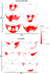
Maps illustrating the significance of local differences in (a) circumferential and (b) radial stretch between each TAV leaflet type and their corresponding BAV leaflet types, both fused and non-fused. Herein, color is proportional to log10(p), where p is the p-value resulting from the pointwise t-test. Regions with lower p (i.e. colored more red) show more consistent differences in stretch.

References

    1. Aggarwal A, Ferrari G, Joyce E, Daniels MJ, Sainger R, Gorman JH, Gorman R, and Sacks MS. Architectural trends in the human normal and bicuspid aortic valve leaflet and its relevance to valve disease. Ann. Biomed. Eng 42(5):986–998, 2014. - PMC - PubMed
    1. Aggarwal A, Pouch AM, Lai E, Lesicko J, Yushkevich PA, Gorman JH, Gorman RC, and Sacks MS. In-vivo heterogeneous functional and residual strains in human aortic valve leaflets. J. Biomech 49(12):2481–2490, 2016. - PMC - PubMed
    1. Ayoub S, Ferrari G, Gorman RC, Gorman JH, Schoen FJ, and Sacks MS. Heart valve biomechanics and underlying mechanobiology. Compr. Physiol 6(4):1743–1780, 2016. - PMC - PubMed
    1. Ayoub S, Lee C-H, Driesbaugh KH, Anselmo W, Hughes CT, Ferrari G, Gorman RC, Gorman JH, and Sacks MS. Regulation of valve interstitial cell homeostasis by mechanical deformation: implications for heart valve disease and surgical repair. J. R. Soc. Interface 14(135):20170580, 2017. - PMC - PubMed
    1. Balachandran K, Sucosky P, Jo H, and Yoganathan AP. Elevated cyclic stretch alters matrix remodeling in aortic valve cusps: implications for degenerative aortic valve disease. Am. J. Physiol.-Heart Circ. Physiol 296(3):H756–H764, 2009. - PubMed