. 2022 Jan 6;29(1):116-130.e7.
doi: 10.1016/j.stem.2021.12.002.

Down-syndrome-induced senescence disrupts the nuclear architecture of neural progenitors

Affiliations

Down-syndrome-induced senescence disrupts the nuclear architecture of neural progenitors

Hiruy S Meharena et al. Cell Stem Cell. .

Abstract

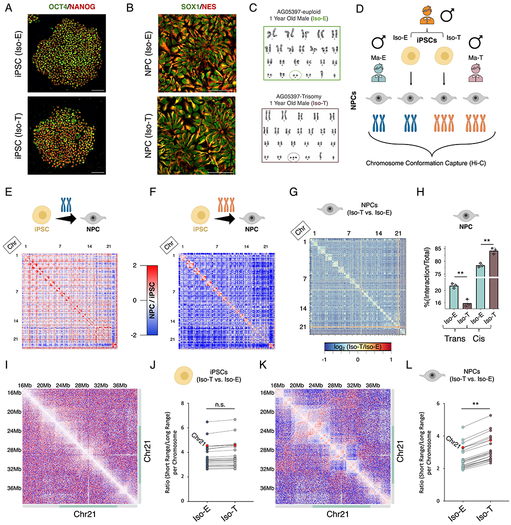
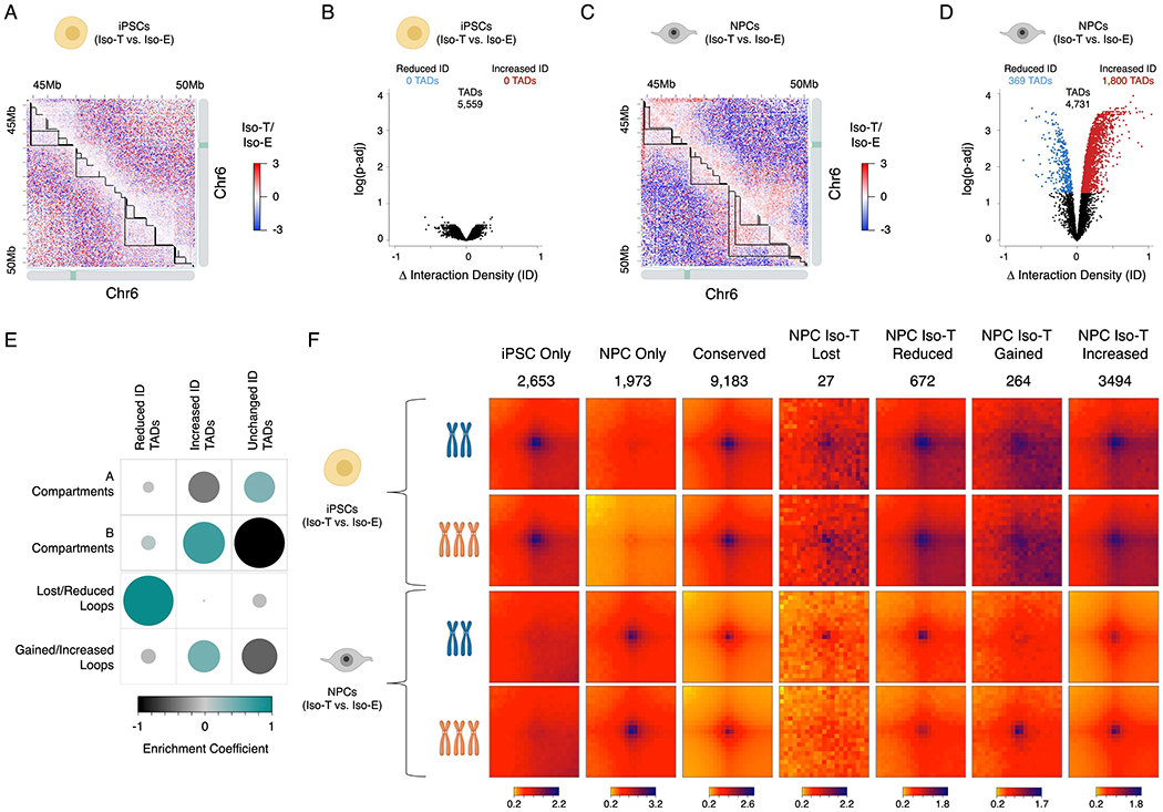
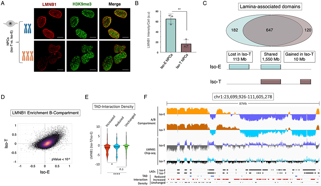
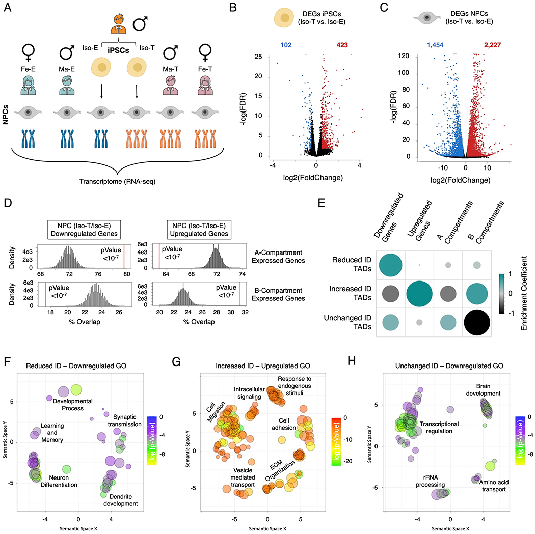
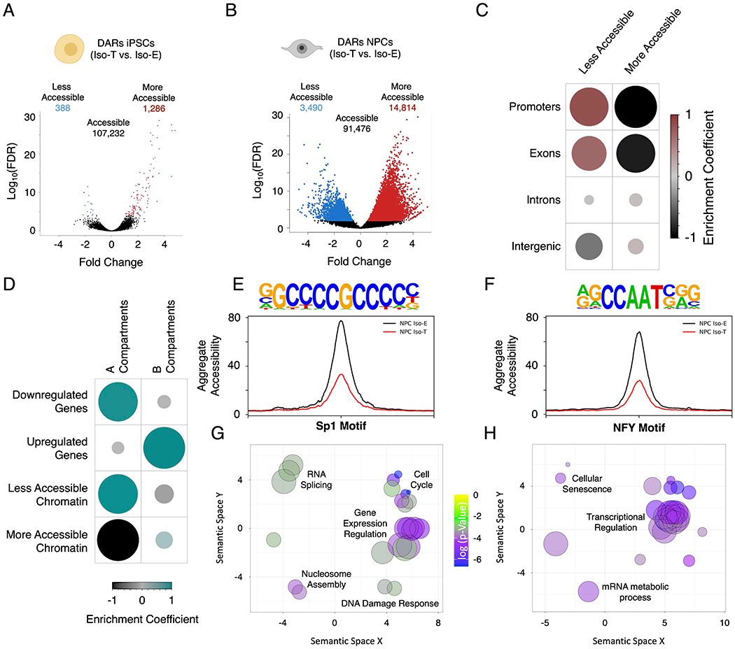
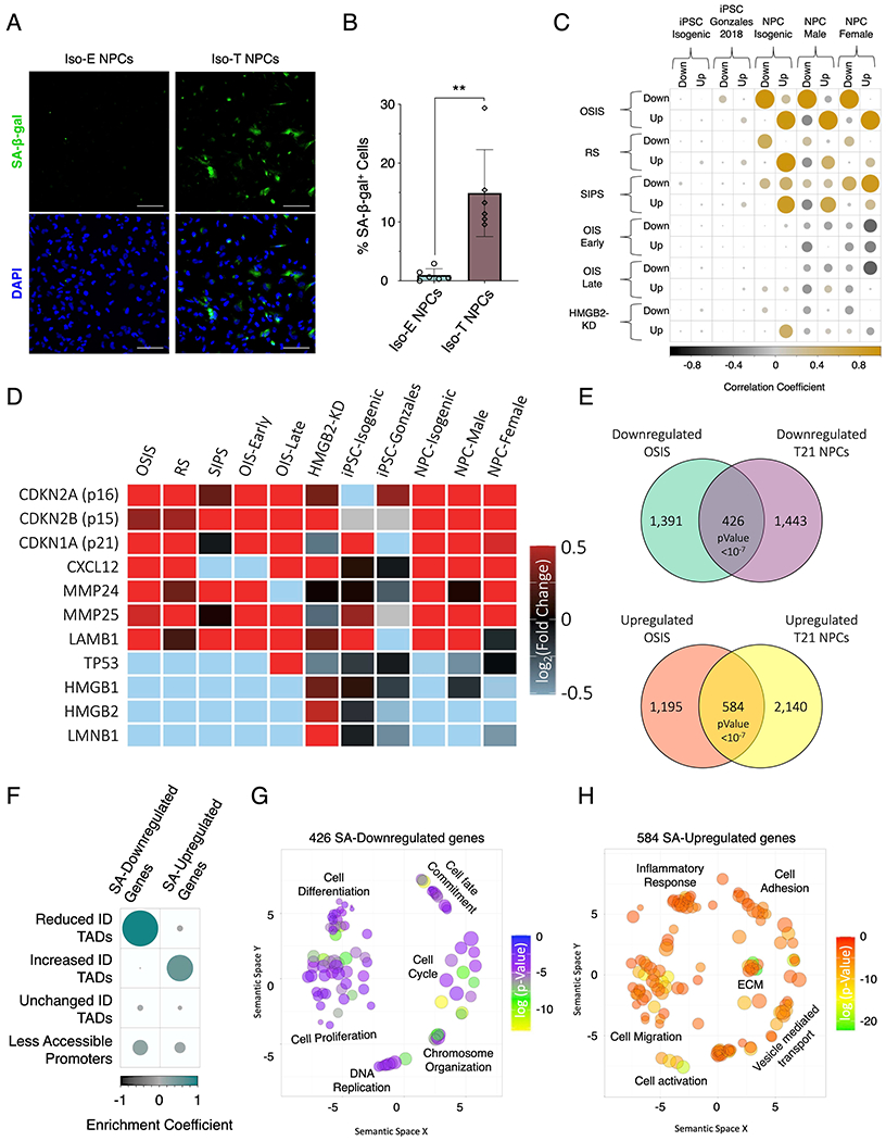
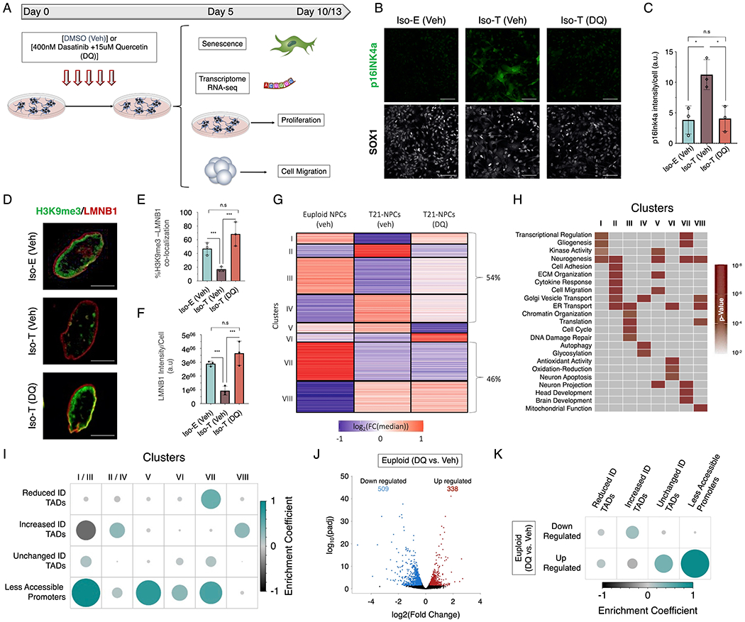
Down syndrome (DS) is a genetic disorder driven by the triplication of chromosome 21 (T21) and characterized by a wide range of neurodevelopmental and physical disabilities. Transcriptomic analysis of tissue samples from individuals with DS has revealed that T21 induces a genome-wide transcriptional disruption. However, the consequences of T21 on the nuclear architecture and its interplay with the transcriptome remain unknown. In this study, we find that unlike human induced pluripotent stem cells (iPSCs), iPSC-derived neural progenitor cells (NPCs) exhibit genome-wide "chromosomal introversion," disruption of lamina-associated domains, and global chromatin accessibility changes in response to T21, consistent with the transcriptional and nuclear architecture changes characteristic of senescent cells. Treatment of T21-harboring NPCs with senolytic drugs alleviates the transcriptional, molecular, and cellular dysfunctions associated with DS. Our findings provide a mechanistic link between T21 and global transcriptional disruption and indicate that senescence-associated phenotypes may play a key role in the neurodevelopmental pathogenesis of DS.

Keywords: 3D-genome; ATAC-seq; Down syndrome; Hi-C; RNA-seq; aneuploidy; epigenome; lamina-associated domains (LADs); senescence; senolytic drugs; transcriptome.


Conflict of interest statement

Declaration of interests The authors declare no competing interests.

Figures

Figure 1 –
Figure 1 –. T21 induces genome-wide chromosomal introversion in NPCs but not iPSCs.
Immunofluorescence staining of (A) iPSCs (OCT4 (green), NANOG (red)) and (B) NPCs (SOX1 (green), Nestin (red)) from the isogenic pair (Iso-E (top) and Iso-T (bottom) as well as (C) respective G-band karyotyping of NPCs. (D) Schematic of experimental design to assess the 3D-genome of the isogenic pair iPSCs and NPCs as well as the euploid (Ma-E) and trisomic (Ma-T) male NPCs. (E-H) Comparative Hi-C analysis of Iso-E and Iso-T iPSCs and NPCs. Genome-wide differential (NPC/iPSC) Hi-C interaction maps for Iso-E (E) and Iso-T (F). (G) Genome-wide differential (Iso-T/Iso-E) Hi-C interaction maps for NPCs. (H) Percent distribution of trans- and cis-chromosomal interactions for the isogenic pair of NPCs. (I-L) Representative images of chromosome 21 (chr21) differential cis-chromosomal interaction maps for the isogenic pair of iPSCs (I) and NPCs (K). Dot plots representing the ratio of short-range (<1Mb) to long-range (>1Mb) interactions for the isogenic pair of iPSCs (J) and NPCs (L). Each dot represents a chromosome and chr21 is represented as a red dot. Wilcoxon rank-sum test shows that euploid and trisomic iPSCs were not statistically different (p-value=0.536) whereas NPCs show significantly increased short range interactions in the trisomic NPCs (p-value=0.004).
Figure 2 –
Figure 2 –. T21 induced reorganization of TADs and loops are segregated into A/B compartments.
Representative images of differential Hi-C interaction maps (Iso-T/Iso-E) of a region on chromosome 6 (chr6) in the isogenic pair of iPSCs (A) and NPCs (C). Volcano plot of differential interaction density (ID) within TADs in the isogenic pair of iPSCs (B) and NPCs (D). (E) Randomized permutation test analysis between the differential interaction density (ID) TADs with A/B compartments and differential chromosomal loops in NPCs. Color intensity (positive correlations are displayed in green and negative correlations in black) and the size of the circle are proportional to the enrichment coefficients of A/B compartments with differentially interacting TADs identified as a consequence of T21 in NPCs. (F) Aggregate Peak Analysis (APA) analysis of loops uniquely identified in iPSCs (top rows), NPCs (bottom rows) in both Iso-E and Iso-T.
Figure 3 –
Figure 3 –. T21 induced disruption of lamina associated domains (LADs) is associated with chromosomal introversion.
(A) Immunofluorescence of LMNB1 (red) and H3K9me3 (green) for the isogenic pair to assess LADs and heterochromatin distribution in NPCs. (B) Quantification of LMNB1 staining intensity in the isogenic pair of NPCs. Each dot on the histogram represents a replicate experiment of ~440 total nuclei analyzed for the isogenic pair of NPCs. (C) Venn diagram of LAD overlap between the euploid (Iso-E) and T21 (Iso-T) NPCs (intersect of replicates); bottom, schematic representation of genome coverage (base pairs) by LADs gained or lost as a consequence of T21 relative to euploid. (D) Scatter plot of LMNB1 enrichment over input in Iso-E (x-axis) and Iso-T (y-axis) of the genomic region in the B-compartment. (E) Violin plot of fold change of LMNB1 (Iso-T/Iso-E) of the differential (increased-ID (red), reduced-ID (blue) and unchanged-ID (green)) interaction density TADs. (F) IGV plot of A-compartment (Iso-E (yellow) and Iso-T (orange)) and B-compartment (Iso-E (blue) and Iso-T (light-blue)), LMNB1 ChIP-seq of Iso-E (blue) and Iso-T (light-blue), as well as the location of LADs (black) and the differentially interacting TADs (reduced-ID (blue), increased-ID (red) and unchanged-ID (gray)) an 87Mb region of chromosome 1.
Figure 4 –
Figure 4 –. T21 DEGs are distinctly separated into the A/B compartments in NPCs.
(A) Schematic of experimental design to assess the transcriptional consequences of T21 on the isogenic pair (Iso-E and Iso-T) of iPSCs and NPCs as well as NPCs derived from euploid (female (Fe-E) and male Ma-E)) and trisomic (female (Fe-T) and male (Ma-T)) individuals. (B, C) Volcano plots of the differentially expressed genes (DEGs) identified in the isogenic pair of iPSCs (B) and NPCs. (C). (D) Randomized permutation test between the downregulated and upregulated DEGs induced by T21 in NPCs with A/B compartments. Histogram represents the range of expected overlap percentage and the red line represents the observed percentage overlap. (E) Corrplot of the randomized permutation test between TADs with varying interaction densities (ID) with DEGs and A/B compartments. Color intensity (positive correlations are displayed in green and negative correlations in black) and the size of the circle are proportional to the enrichment coefficients. (F-H) Gene ontology visualized in semantic similarity-based scatterplots for downregulated genes associated with reduced-ID TADs (F), upregulated genes associated with increased-ID TADs (G), and downregulated genes localized within unchanged-ID TADs (H).
Figure 5 –
Figure 5 –. T21-induced genome-wide disruption of chromatin state in NPCs is associated with transcriptional downregulation.
(A, B) Volcano plots of differentially accessible regions (DARs) identified by ATAC-sequencing in the isogenic pair of iPSCs (A) and NPCs (B). (C) Enrichment heatmap of the randomized permutation test between the DARs on promoters, exons, introns and intergenic regions with accessible regions genome-wide. Color intensity (positive correlations are displayed in red and negative correlations in black) and the size of the circle are proportional to the enrichment coefficients. (D) Enrichment heatmap of the randomized permutation test between the A/B compartments and DEGs and DARs. Color intensity (positive correlations are displayed in green and negative correlations in black) and the size of the circle are proportional to the enrichment coefficients of the specific genetic features with the A/B compartments. (E, F) Aggregate plots of less accessible regions harboring SP1 (E) and NFY (F) motifs. (G, H) Gene ontology visualized in semantic similarity-based scatterplots for downregulated genes with less accessible promoters harboring SP1 (G) and NFY (H) motifs.
Figure 6 –
Figure 6 –. T21 induces senescence in NPCs
(A) Senescence associate β-galactosidase staining (SA-β-gal, green) marking senescent NPCs in euploid NPCs (left) and T21 (right). (B) Quantification of SA-β-gal staining intensity in the isogenic pair of NPCs. Each dot on the histogram represents a replicate experiment of ~200 cells analyzed for the isogenic pair of NPCs. (C) Correlation heatmap of the randomized permutation test of DEGs identified in isogenic, male, and female control and T21 iPSCs and NPCs as well as DEGs identified from a previous published iPSC data set (Gonzales), compared to previously published DEGs identified in various modes of senescence induction (oxidative stress induced senescence (OSIS), replicative senescence (RS), stress induced senescence (SIPS), oncogene induced senescence (OIS) at early and late timepoints, as well as senescence entry induced by HMGB2 knock down (HMGB2-KD). Color intensity (positive correlations are displayed in yellow and negative correlations in black) and the size of the circle are proportional to the enrichment coefficients. (D) Gene expression heatmap of senescence marker genes as fold change of T21/E21 or senescent/control. (E) Venn diagram of overlapping downregulated (top) and upregulated (bottom) identified in NPCs harboring T21 and compared to DEGs identified in OSIS induced senescent cells. (F) Enrichment heatmap of the randomized permutation test between DEGs identified in both T21-NPCs and OSIS-induced senescent cells with differential ID TADs as well as reduced promoter-accessibility. Color intensity (positive correlations are displayed in green and negative correlations in black) and the size of the circle are proportional to the enrichment coefficients. (G, H) Gene ontology visualized in semantic similarity-based scatterplots for downregulated (G) and upregulated (H) overlapping genes identified in both T21-NPCs and OSIS-induced senescent cells.
Figure 7 –
Figure 7 –. Senolytic drug alleviates the transcriptional and cellular dysfunctions induced by T21 in NPCs.
(A) Schematic of experimental design to assess the transcriptional and cellular consequences of the senolytic drug (DQ) treatment in isogenic T21 NPCs. (B) Immunofluorescence of p16INK4a (green) in euploid (Iso-E) and trisomic (Iso-T) NPCs (marked by SOX1, white) treated with vehicle or DQ. (C) Quantification of p16INK4a staining intensity in euploid (Iso-E) and trisomic (Iso-T, vehicle or DQ treatment) NPCs. (D) Immunofluorescence of H3K9me3 (green) and LMNB1 (red) in euploid (Iso-E) and trisomic (Iso-T) NPCs treated with vehicle or DQ. (E) Quantification of co-localization between H3K9me3 and LMNB1 in euploid (Iso-E) and trisomic (Iso-T) NPCs and (F) quantification of LMNB1 staining intensity of NPCs treated with vehicle or DQ. Each dot on the histogram represents a replicate and ~200 nuclei were analyzed per condition. (G) K-means clustering of the transcriptomic analysis of euploid (Iso-E) and trisomic (Iso-T) NPCs (treated with vehicle or DQ) identified 8 distinct clusters. Values in heatmap are plotted as the log2 fold change from the median. (H) Gene ontology of biological process associated with the 8 transcriptional clusters identified in response to senolytic drug treatment. (I) Enrichment plot of the randomized permutation test between the genes identified in the 8 different clusters with differential ID TADs as well as less accessible promoters. Genes from cluster I and III as well clusters II and IV were merged for this analysis. Color intensity (positive correlations are displayed in green and negative correlations in black) and the size of the circle are proportional to the enrichment coefficients. (J) Volcano plots of the differentially expressed genes (DEGs) identified in euploid NPCs treated with vehicle (DMSO) or DQ. (K) Enrichment plot of the randomized permutation test between DEGs identified in DEGs identified after DQ treatment with differential ID TADs as well as reduced promoter-accessibility. Color intensity (positive correlations are displayed in green and negative correlations in black) and the size of the circle are proportional to the enrichment coefficients.

Similar articles

Cited by

References

    1. Abdennur N, and Mirny LA (2020). Cooler: scalable storage for Hi-C data and other genomically labeled arrays. Bioinformatics 36, 311–316. - PMC - PubMed
    1. Aldridge K, Reeves RH, Olson LE, and Richtsmeier JT (2007). Differential effects of trisomy on brain shape and volume in related aneuploid mouse models. Am J Med Genet A 143A, 1060–1070. - PMC - PubMed
    1. Allis CD, and Jenuwein T (2016). The molecular hallmarks of epigenetic control. Nat Rev Genet 17, 487–500. - PubMed
    1. Alspach E, Flanagan KC, Luo X, Ruhland MK, Huang H, Pazolli E, Donlin MJ, Marsh T, Piwnica-Worms D, Monahan J, et al. (2014). p38MAPK plays a crucial role in stromal-mediated tumorigenesis. Cancer Discov 4, 716–729. - PMC - PubMed
    1. Anders S, Pyl PT, and Huber W (2015). HTSeq--a Python framework to work with high-throughput sequencing data. Bioinformatics 31, 166–169. - PMC - PubMed

Publication types