Review
. 2022 Jan 6;185(1):77-94.
doi: 10.1016/j.cell.2021.10.029.

Central nervous system regeneration

Affiliations
Review

Central nervous system regeneration

Supraja G Varadarajan et al. Cell. .

Abstract

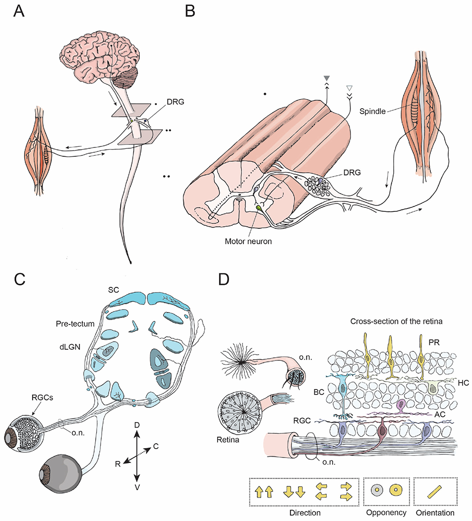
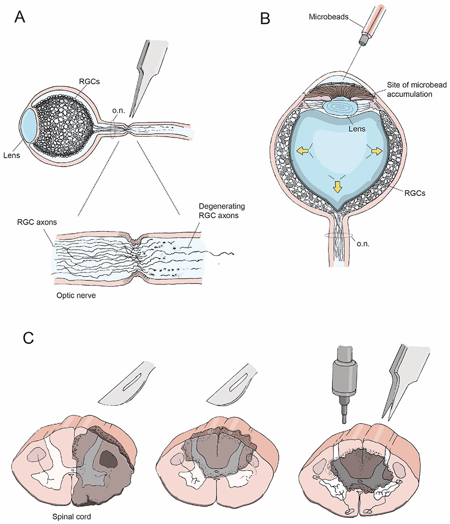
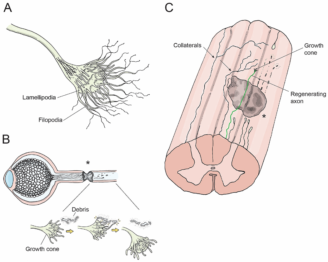
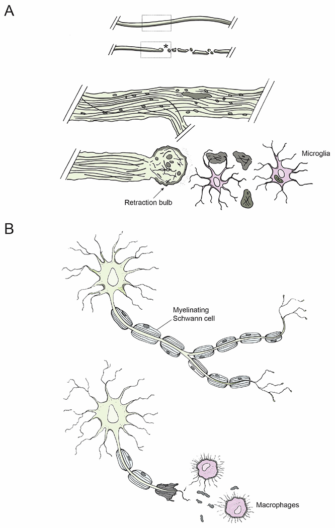
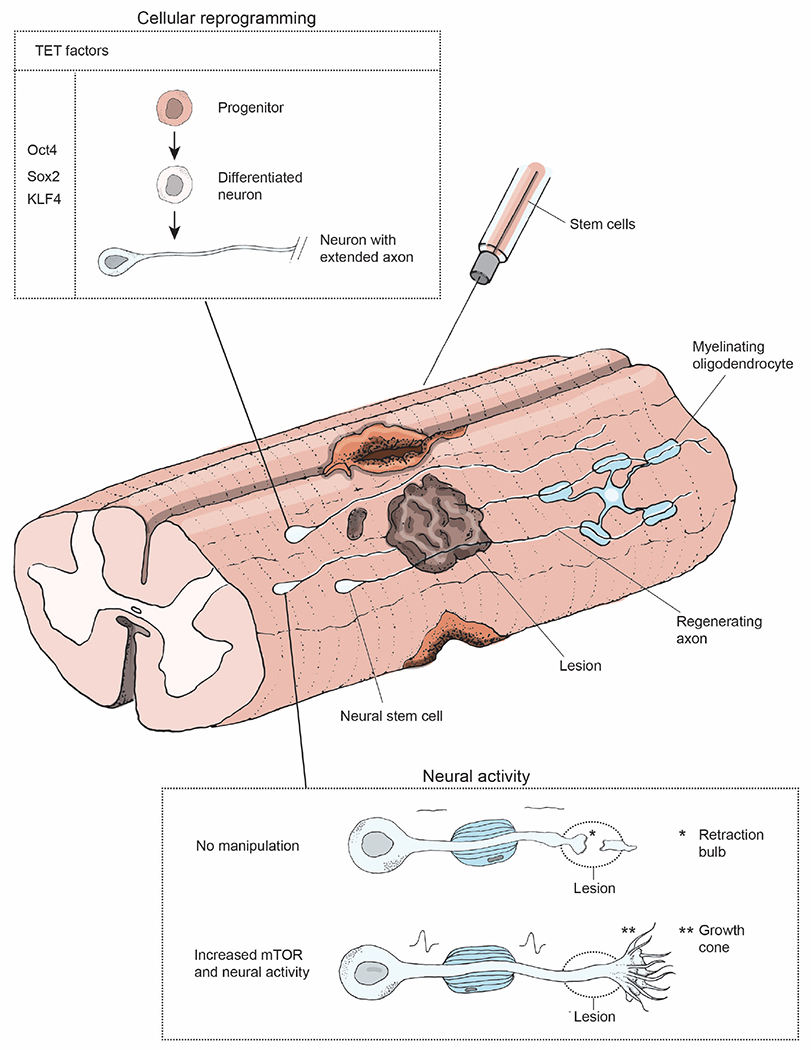
Neurons of the mammalian central nervous system fail to regenerate. Substantial progress has been made toward identifying the cellular and molecular mechanisms that underlie regenerative failure and how altering those pathways can promote cell survival and/or axon regeneration. Here, we summarize those findings while comparing the regenerative process in the central versus the peripheral nervous system. We also highlight studies that advance our understanding of the mechanisms underlying neural degeneration in response to injury, as many of these mechanisms represent primary targets for restoring functional neural circuits.


Conflict of interest statement

Declaration of interests The authors declare no competing interests.

Figures

Figure 1:
Figure 1:. Model systems for studying CNS regeneration: spinal system and visual system.
(A) The brain and the spinal cord exchange information via descending projections from the brain to the spinal cord (arrow), and ascending projections. The spinal cord receives sensory input from external stimuli via the dorsal root ganglia (DRG) neurons. Output signals from the brain and the spinal cord are relayed to the muscles via motor neurons and their peripheral projections. (B) A longitudinal view of the spinal cord showing sensory inputs, interneurons, and motor outputs. The spinothalamic tract is comprised of ascending projections from interneurons (gray, closed arrowhead), whereas the corticospinal tract consists of descending projections from the brain to the spinal cord (gray, open arrowhead). The DRG neurons have central and peripheral axonal branches. (C) The visual system includes retinal ganglion cells (RGCs, arrow), the neurons that relay sensory input from the eye, their axons that form the optic nerve (o.n.) and their projections to target regions within the brain including the dorsal lateral geniculate nucleus (dLGN) and the superior colliculus (SC) (blue). (D) Cross-section of the retina and the optic nerve. The retina consists of five types of neural cells: photoreceptors, i.e. rods and cones (PR, yellow), bipolar cells (BC, blue), horizontal cells (HC, green), amacrine cells (AC, pink) and ~40 different types of retinal ganglion cells (RGC, brown, purple). RGCs are the only neurons that send projections from the retina into the optic nerve and have various receptive field properties as indicated by the yellow arrows and circles.
Figure 2:
Figure 2:. CNS injury models
(A) Optic nerve crush injury showing axon degeneration distal to the injury site (B) Bead-induced mouse model of glaucoma, wherein microbeads injected into the eye increase intraocular pressure (arrows) and mimic the degenerative effects of glaucoma (C) Coronal views of the spinal cord depicting lesion sites (gray) following unilateral transection (left), dorsal bilateral hemisection (middle), and a contusion or crush injury (right).
Figure 3:
Figure 3:. Axon growth following injury
(A) Growth cone, i.e. the leading edge of an axon. The fingerlike protrusions are filiopodia and lamellipodia that are composed of actin filaments and are crucial for the growth cone’s ability to grow towards or away from environmental cues. (B) Axons regenerating following an optic nerve crush injury. Growth cones at the leading edge of regenerating axons grow past the lesion site (asterisk), interact with microglia/axonal-debris/guidance cues, and accordingly alter their direction of growth. (C) Longitudinal view of the spinal cord showing a lesion site (gray), distal processes of injured axons degenerating (dotted lines), a regenerating axon extending a growth cone distal to the lesion site (color), and two spared axons extending collaterals circumventing the lesion site and extending to target neurons distal to the lesion (arrows).
Figure 4:
Figure 4:. Axon degeneration and immune response to injury
(A) Degenerative mechanisms following injury: An intact axon before injury; an injured axon undergoing Wallerian degeneration distal to the lesion site (asterisk). Dotted regions of the intact and injured axons are shown magnified below: retraction bulb (arrow) sealing the axolemma at the proximal end of an injured axon and microglia (pink) clearing axonal debris. (B) Injury in the PNS: Schwann cells in the PNS myelinate regenerating axons (top); macrophages phagocytose axonal debris (bottom, pink).
Figure 5:
Figure 5:. Clinically relevant therapeutic strategies
The most promising therapeutic strategies to promote re-connectivity of neural circuits are illustrated. Stem cells can be utilized to promote functional recovery following injury by neural stem cell grafts or by overexpressing TET factors (Oct4, Sox2, and KLF4)(Kumamaru et al., 2019; Lu et al., 2020) to reprogram cells to a development-like state that encourages axon regrowth. Upregulation of mTOR signaling and neuronal activity can promote axon regeneration past the lesion site(Lim et al., 2016), while voltage-gated potassium channel blocker can be utilized to promote myelin reformation in regenerating axons (Bei et al., 2016).

References

    1. Adams KL, and Gallo V (2018). The diversity and disparity of the glial scar. Nat. Neurosci 21, 9–15. - PMC - PubMed
    1. Ambacher KK, Pitzul KB, Karajgikar M, Hamilton A, Ferguson SS, and Cregan SP (2012). The JNK- and AKT/GSK3β- signaling pathways converge to regulate Puma induction and neuronal apoptosis induced by trophic factor deprivation. PLoS One 7, e46885. - PMC - PubMed
    1. Ambros V, and Horvitz HR (1984). Heterochronic mutants of the nematode Caenorhabditis elegans. Science 226, 409–416. - PubMed
    1. Anderson MA, Burda JE, Ren Y, Ao Y, O’Shea TM, Kawaguchi R, Coppola G, Khakh BS, Deming TJ, and Sofroniew MV (2016). Astrocyte scar formation aids central nervous system axon regeneration. Nature 532, 195–200. - PMC - PubMed
    1. Andrews MR, Czvitkovich S, Dassie E, Vogelaar CF, Faissner A, Blits B, Gage FH, ffrench-Constant C, and Fawcett JW (2009). Alpha9 integrin promotes neurite outgrowth on tenascin-C and enhances sensory axon regeneration. J. Neurosci 29, 5546–5557. - PMC - PubMed

Publication types

LinkOut - more resources