. 2022 Jan 7;20(1):9.
doi: 10.1186/s12915-021-01220-z.

Evolutionary analyses of the gasdermin family suggest conserved roles in infection response despite loss of pore-forming functionality

Affiliations

Evolutionary analyses of the gasdermin family suggest conserved roles in infection response despite loss of pore-forming functionality

Diego Angosto-Bazarra et al. BMC Biol. .

Abstract

Background: Gasdermins are ancient (>500million-years-ago) proteins, constituting a family of pore-forming proteins that allow the release of intracellular content including proinflammatory cytokines. Despite their importance in the immune response, and although gasdermin and gasdermin-like genes have been identified across a wide range of animal and non-animal species, there is limited information about the evolutionary history of the gasdermin family, and their functional roles after infection. In this study, we assess the lytic functions of different gasdermins across Metazoa species, and use a mouse model of sepsis to evaluate the expression of the different gasdermins during infection.

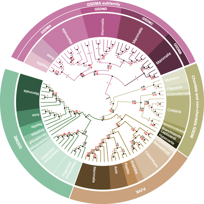
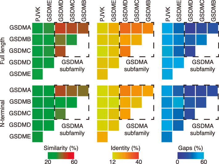
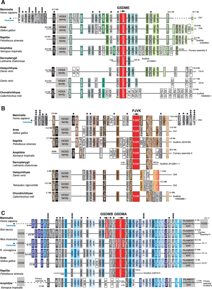
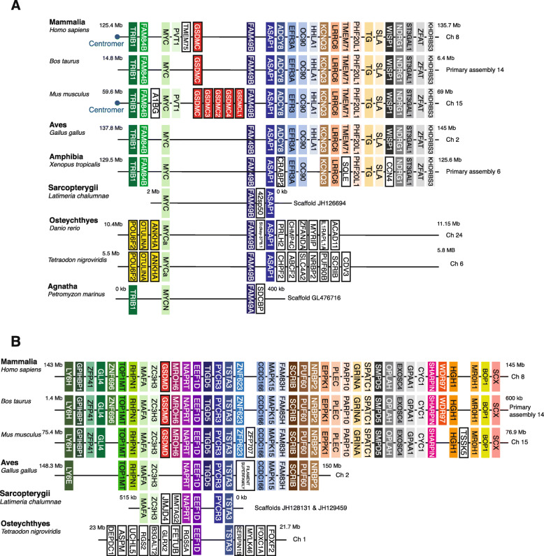
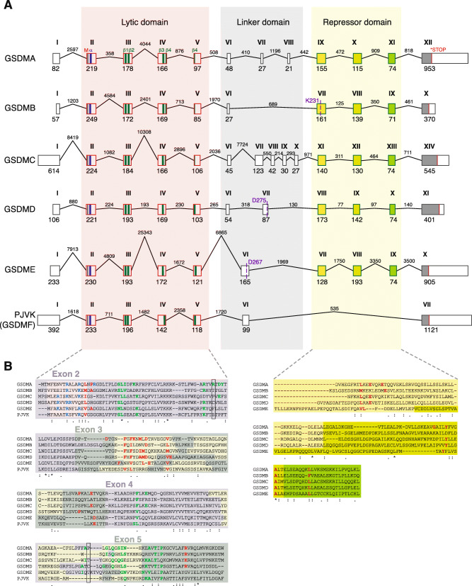
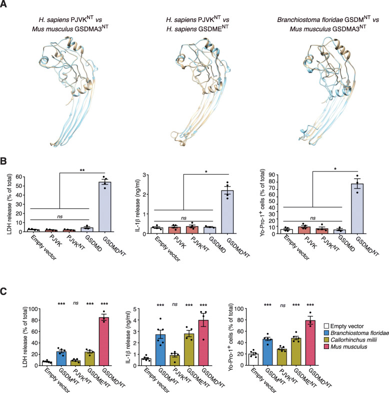
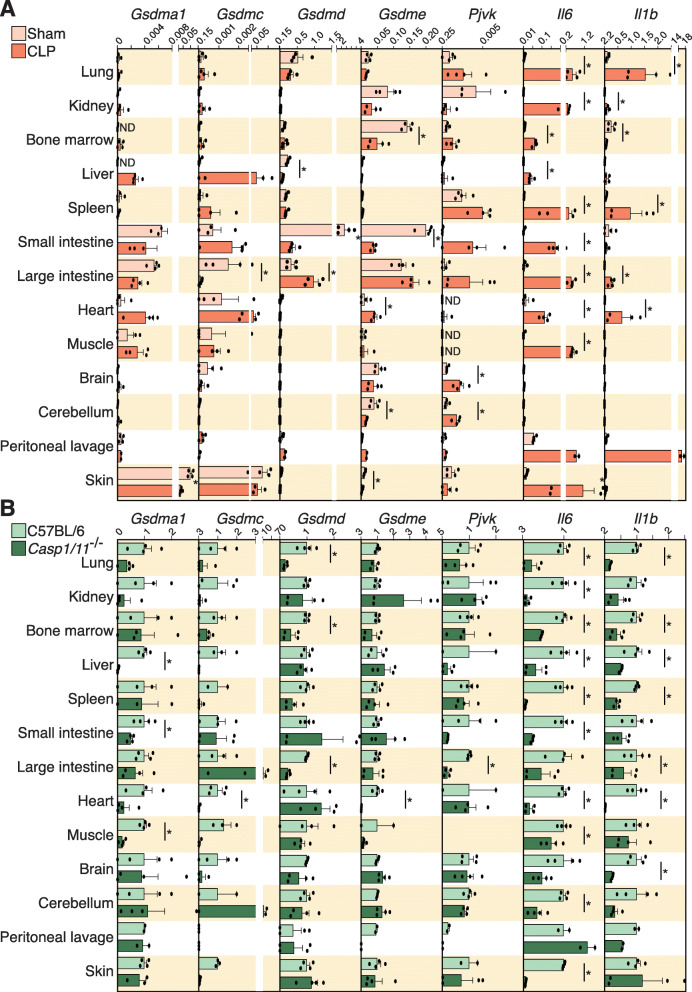
Results: We show that the majority of gasdermin family members from distantly related animal clades are pore-forming, in line with the function of the ancestral proto-gasdermin and gasdermin-like proteins of Bacteria. We demonstrate the first expansion of this family occurred through a duplication of the ancestral gasdermin gene which formed gasdermin E and pejvakin prior to the divergence of cartilaginous fish and bony fish ~475 mya. We show that pejvakin from cartilaginous fish and mammals lost the pore-forming functionality and thus its role in cell lysis. We describe that the pore-forming gasdermin A formed ~320 mya as a duplication of gasdermin E prior to the divergence of the Sauropsida clade (the ancestral lineage of reptiles, turtles, and birds) and the Synapsid clade (the ancestral lineage of mammals). We then demonstrate that the gasdermin A gene duplicated to form the rest of the gasdermin family including gasdermins B, C, and D: pore-forming proteins that present a high variation of the exons in the linker sequence, which in turn allows for diverse activation pathways. Finally, we describe expression of murine gasdermin family members in different tissues in a mouse sepsis model, indicating function during infection response.

Conclusions: In this study we explored the evolutionary history of the gasdermin proteins in animals and demonstrated that the pore-formation functionality has been conserved from the ancient proto-gasdermin protein. We also showed that one gasdermin family member, pejvakin, lost its pore-forming functionality, but that all gasdermin family members, including pejvakin, likely retained a role in inflammation and the physiological response to infection.

Keywords: Evolution; Gasdermin; Infection; Pejvakin; Pyroptosis; Sepsis.


Conflict of interest statement

The authors declare that they have no competing interests.

Figures

Fig. 1
Fig. 1
Phylogeny of the gasdermin family. Phylogenetic tree inferred from amino acid sequence data using the Bayesian (black numbers) and maximum likelihood (red numbers) methods showing divergence of full-length gasdermins in mammals, birds, reptiles, amphibians, fish, jawless Chordata, and non-Chordata. Black numbers show the posterior distribution of the Bayesian method and red numbers show the bootstrap analysis of the maximum likelihood analysis
Fig. 2
Fig. 2
Human gasdermin sequence similarity. Heat maps showing the percentage of similarity (left), identity (middle), and gaps (right) between the full-length gasdermin sequences (top) and their respective N-terminal domain (bottom). GSDMA subfamily is highlighted with a dashed line box and presents the higher similarity
Fig. 3
Fig. 3
Synteny analysis of the gasdermin E, pejvakin, and gasdermin A/B loci from different species. Schematic diagrams showing the conservation of synteny in the gsdme loci A, pjvk loci B, and gsdma/b loci C in Mammalia (Homo sapiens, Mus musculus, Rattus norvegicus, Bos taurus), Aves (Gallus gallus), Reptilia (Pediscus sinensis), Amphibia (Xenopus tropicalis), and fish (Osteychthyes: Latimeria chalumnae, Danio rerio, Tetraodon nigroviridis, and Chondrichthyes: Callorhinchus milii). In B, for Danio rerio, the synteny of the genes surrounding the mammalian pjvk is presented to illustrate that fish pjvk is not located in that part of the genome. In C, amphibian synteny of genes surrounding mammalian gsdma gene is shown to illustrate that no gsdma is present. Conserved genes are indicated in different colors and the direction of gasdermin gene transcription is indicated with arrows. The position of the centromeres is indicated with a blue dot. An asterisk denotes genes from the same family found in two of the genomic environments (gsdme/pjvk, gsdme/gsdma, or pjvk/gsdma); three asterisks denote genes from the same family found in the three genomic environments
Fig. 4
Fig. 4
Synteny analysis of the gasdermin C and D loci from different species. Schematic diagrams showing the conserved synteny in the gsdmc loci (A) and gsdmd loci (B) in Mammalia (Homo sapiens, Mus musculus, Bos taurus). The synteny of the genes surrounding the mammalian gsdmc/d is shown in Aves (Gallus gallus), Amphibian (Xenopus tropicalis), Fish (Latimeria chalumnae, Danio rerio, Tetraodon nigroviridis), and Agnatha (Petromyzon marinus) to illustrate that no gsdmc or gsdmd is present in these species. Conserved genes are indicated in different colors, and the direction of gasdermin gene transcription is indicated with arrows. The position of the centromeres is indicated with a blue dot
Fig. 5
Fig. 5
Analysis of the exons and introns present in human gasdermin genes. A Schematic representation of the different exons and introns present in the genomic DNA of the different human gasdermins. The first methionine is indicated with a black line, the stop codon with a red line, the first α-helix of the N-terminal domain is indicated with a blue bar, and the four β-sheets that integrate into membranes are indicated with green bars. The exons of the repressor C-terminal domain are represented with different colors to show the conserved similarity between them. Cleavage sites for granzyme A (K231) in human GSDMB, caspase-1 (D275) in human GSDMD and caspase-3 (D267) in human GSDME are shown. For human gsdmb, the gsdmb-403 splice variant is shown. B Protein alignment of the N-terminal and repressor C-terminal domains with the different exons highlighted. The residues that are part of the α-helix are highlighted in yellow, and the positive residues involved in the lipid binding are presented in blue. The residues involved in the insertion into membranes are presented in green and the residues involved in the oligomerization of different subunits to form the pore are presented in red. The positions of the Cys51 and Cys191 of human GSDMD are shown by a black box. In the C-terminal, the residues presented in red are important for the auto-inhibition
Fig. 6
Fig. 6
Full-length or N-terminal pejvakin expression does not induce pyroptosis. A Structural alignment of the molecular models of mouse GSDMA3NT (6CB8) (brown) with the model for the template generated human PJVKNT (cyan) (left panel). Structural alignment of human GSDMENT (brown) with human PJVKNT (Cyan) both generated from the mouse GSDMA3NT template-based model (middle panel). Structural alignment of mouse GSDMA3NT (6CB8) (brown) with the template-based model generated for jawless Chordate lancelet fish (Branchiostoma floridae) GSDMNT (Cyan) (right panel). B Measurement of LDH release, IL-1β release, and Yo-Pro-1+ uptake in HEK293 cells that constitutively express the mature form of human IL-1β and transiently express human full length PJVK, PJVKNT, full length GSDMD, or GSDMDNT. C Measurement of LDH release, IL-1β release, and Yo-Pro-1+ uptake in HEK293 cells that constitutively express the mature form of human IL-1β and transiently express lancelet GSDMNT (blue), shark PJVKNT, or GSDMENT (dark yellow) or mouse GSDMDNT (red). In B and C, each point represents an individual assay with at least n=3 to 6 different assays. *p<0.05, **p<0.01, and ***p<0.001; Kruskal-Wallis test with Dunn’s multiple comparison post-test
Fig. 7.
Fig. 7.
Gene expression of gasdermin genes in different mouse tissues during sepsis. A Relative expression of mouse gsdma1, gsdmc, gsdmd, gsdme, pjvk, il6, and il1b genes analyzed by quantitative PCR from different tissues of sham operated mice (yellow) and septic mice (CLP, in orange). B Fold change of the expression of mouse gsdma1, gsdmc, gsdmd, gsdme, pjvk, il6, and il1b genes analyzed by quantitative PCR from different tissues of septic wild type (C57BL/6, in light green) versus Casp1/11-/- mice (dark green); Casp1/11-/- expression was relative to wild type expression. Each point represents an independent sample from n= 2 mice. Mann-Whitney test for A and B, *p<0.05

References

    1. Saeki N, Kuwahara Y, Sasaki H, Satoh H, Shiroishi T. Gasdermin (Gsdm) localizing to mouse chromosome 11 is predominantly expressed in upper gastroiatestinal tract but significantly suppressed in human gastric cancer cells. Mamm Genome. 2000;11(9):718–724. doi: 10.1007/s003350010138. - DOI - PubMed
    1. Tamura M, Tanaka S, Fujii T, Aoki A, Komiyama H, Ezawa K, Sumiyama K, Sagai T, Shiroishi T. Members of a novel gene family, Gsdm, are expressed exclusively in the epithelium of the skin and gastrointestinal tract in a highly tissue-specific manner. Genomics. 2007;89(5):618–629. doi: 10.1016/j.ygeno.2007.01.003. - DOI - PubMed
    1. Kovacs SB, Miao EA. Gasdermins: Effectors of Pyroptosis. Trends Cell Biol. 2017;27(9):673–684. doi: 10.1016/j.tcb.2017.05.005. - DOI - PMC - PubMed
    1. Broz P, Pelegrín P, Shao F. The gasdermins, a protein family executing cell death and inflammation. Nat Rev Immunol. 2020;20(3):143–157. doi: 10.1038/s41577-019-0228-2. - DOI - PubMed
    1. Ding J, Wang K, Liu W, She Y, Sun Q, Shi J, Sun H, Wang DC, Shao F. Pore-forming activity and structural autoinhibition of the gasdermin family. Nature. 2016;535(7610):111–116. doi: 10.1038/nature18590. - DOI - PubMed

Publication types