. 2022 Feb 1;208(3):732-744.
doi: 10.4049/jimmunol.2100184. Epub 2022 Jan 7.

Siglec-F Promotes IL-33-Induced Cytokine Release from Bone Marrow-Derived Eosinophils Independently of the ITIM and ITIM-like Motif Phosphorylation

Affiliations

Siglec-F Promotes IL-33-Induced Cytokine Release from Bone Marrow-Derived Eosinophils Independently of the ITIM and ITIM-like Motif Phosphorylation

Stefanie Westermann et al. J Immunol. .

Abstract

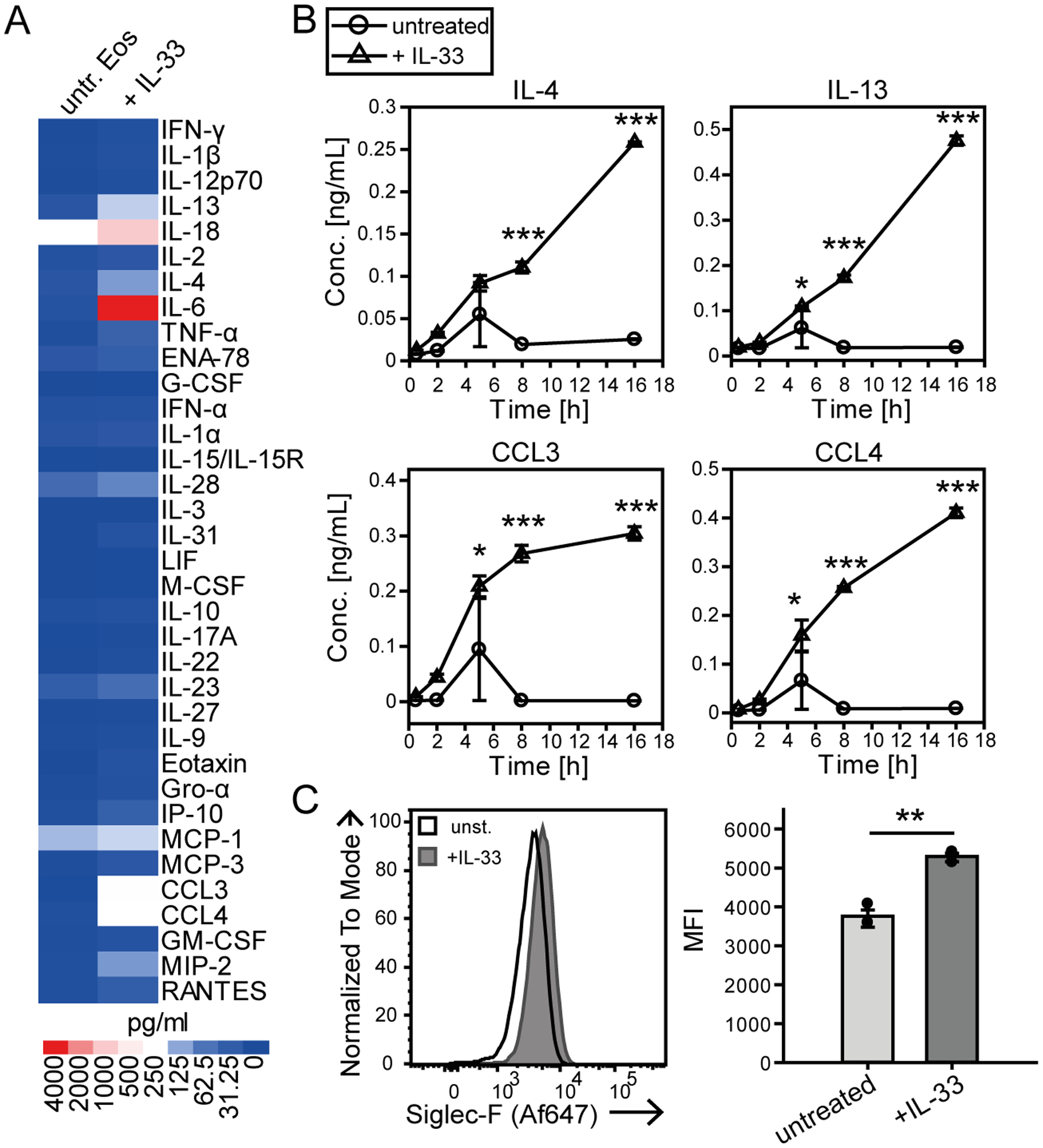
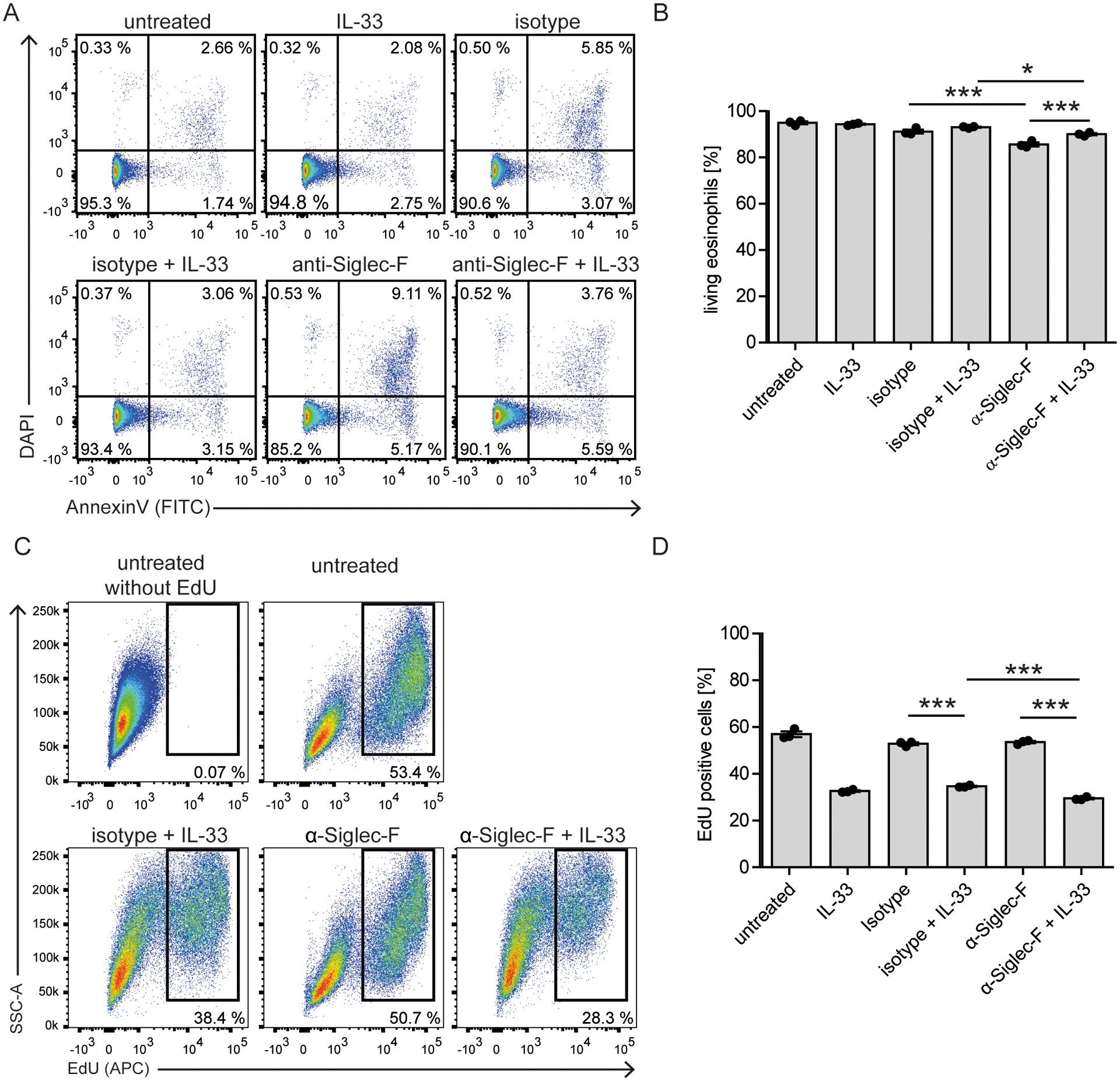
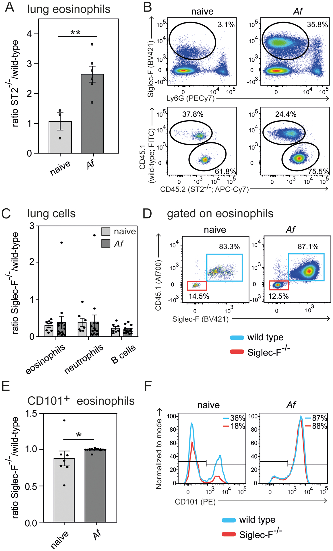
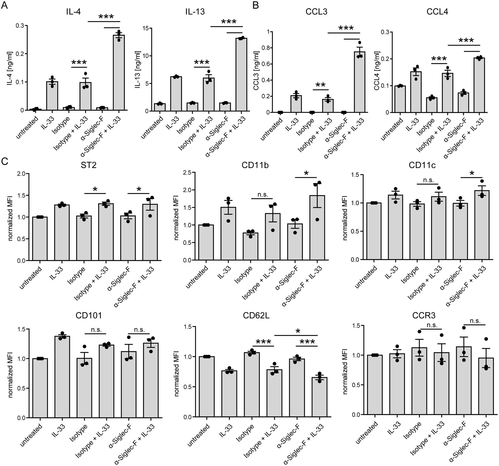
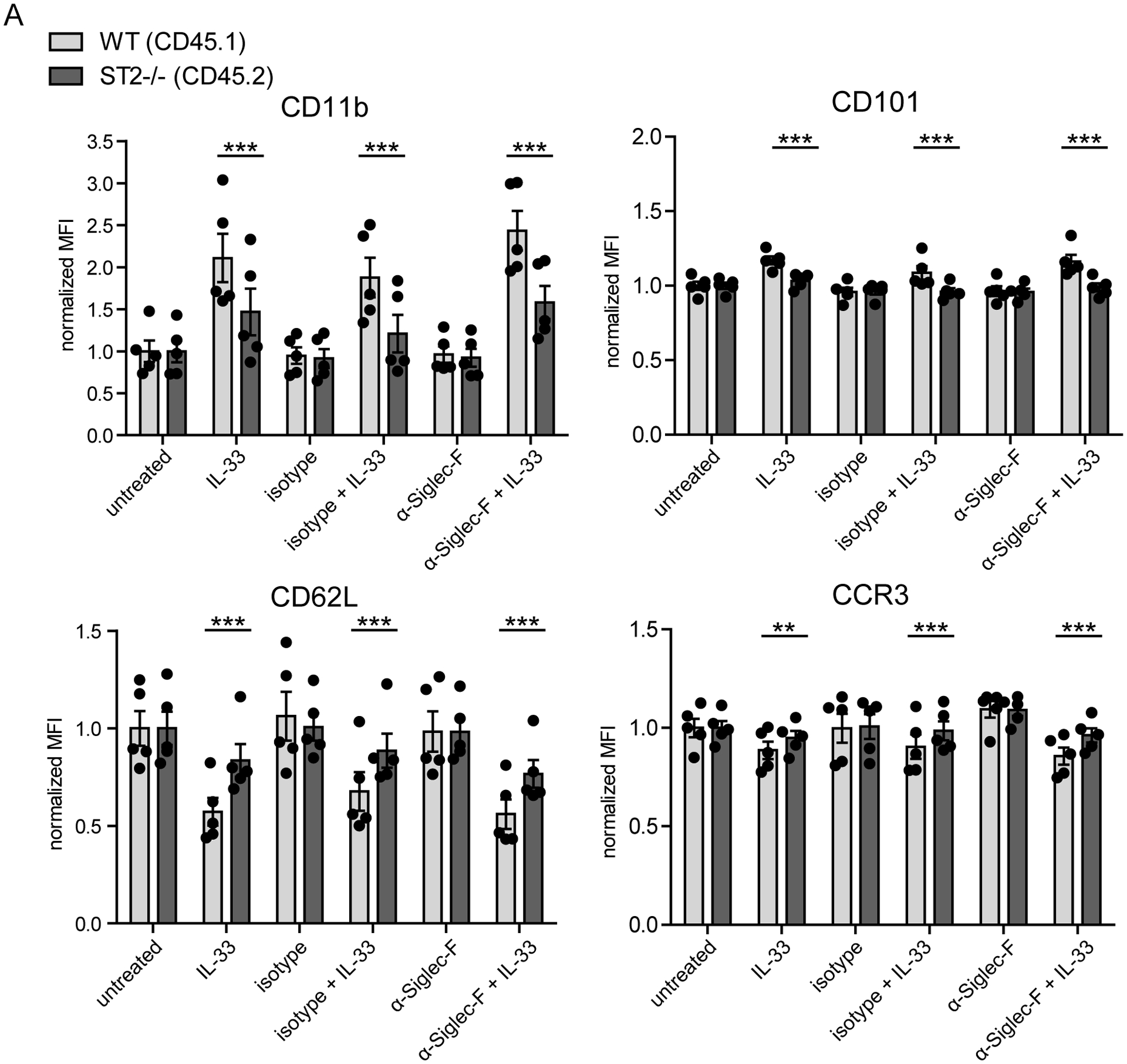
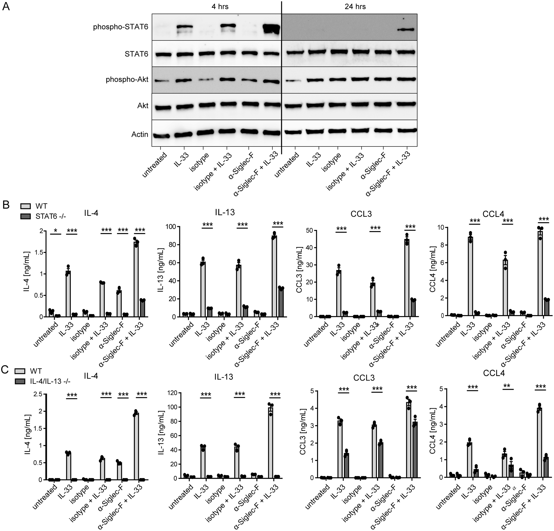
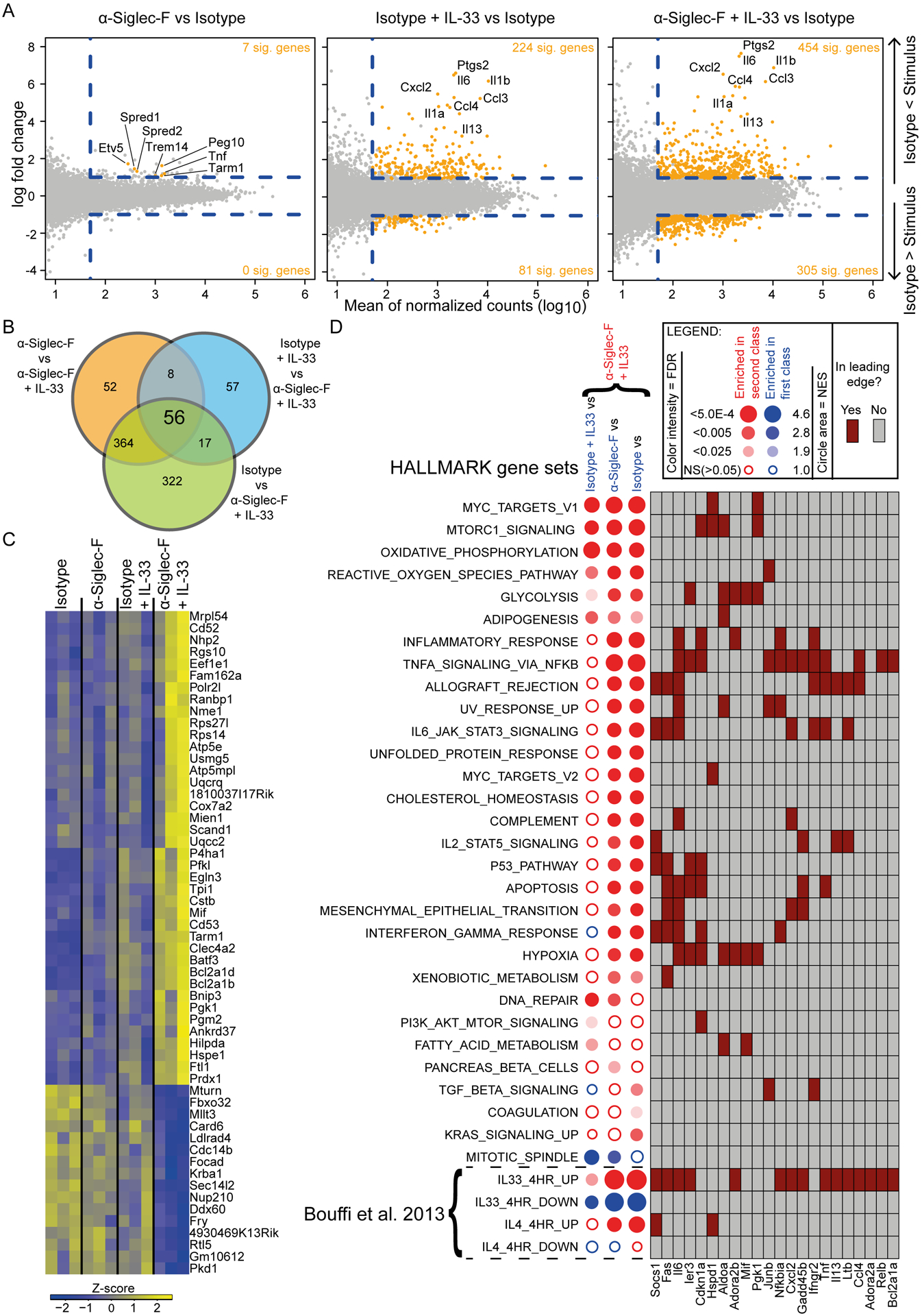
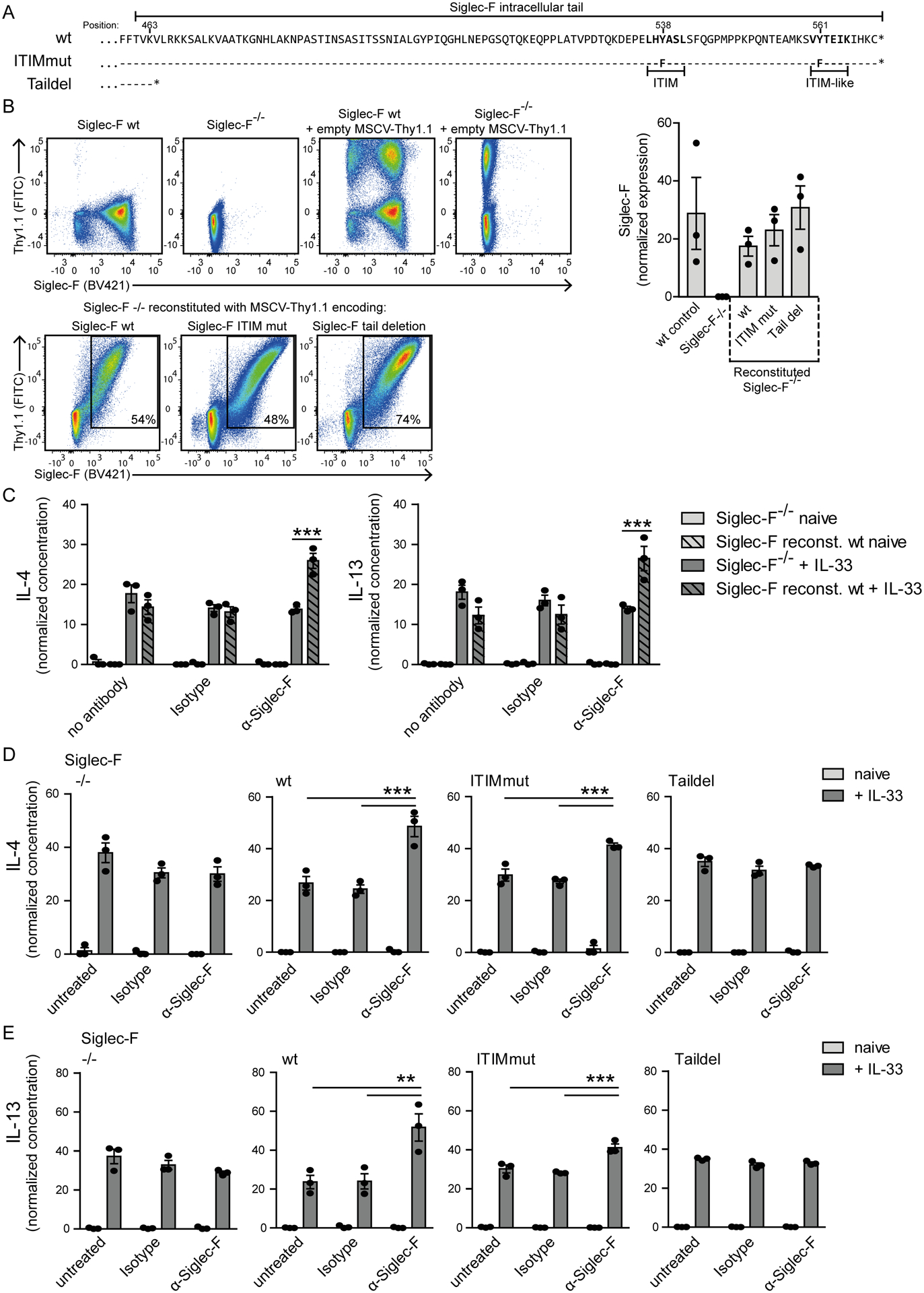
Eosinophils are potent innate effector cells associated mainly with type 2 immune responses elicited by helminths and allergens. Their activity needs to be tightly controlled to prevent severe inflammation and tissue damage. Eosinophil degranulation and secretion of inflammatory effector molecules, including cytokines, chemokines, and lipid mediators, can be regulated by activating and inhibitory receptors on the cell surface. In this study, we investigated the modulation of proliferation, apoptosis, gene expression, and cytokine/chemokine secretion from IL-33-activated Mus musculus eosinophils on cross-linking of the transmembrane receptor Sialic acid-binding Ig-like lectin F (Siglec-F). Siglec-F contains an ITIM plus an ITIM-like motif in its intracellular tail and is mainly regarded as an inhibitory and apoptosis-inducing receptor. In vitro costimulation of bone marrow-derived eosinophils with anti-Siglec-F and IL-33 compared with treatment with either alone led to enhanced STAT6 phosphorylation, stronger induction of hypoxia/glycolysis-related proinflammatory genes, and elevated secretion of type 2 cytokines (IL-4, IL-13) and chemokines (CCL3, CCL4) with only minor effects on proliferation and apoptosis. Using a competitive mixed bone marrow chimera approach with wild-type and Siglec-F-deficient eosinophils, we observed no evidence for Siglec-F-regulated inhibition of Aspergillus fumigatus-elicited lung eosinophilia. Truncation of the Siglec-F cytoplasmic tail, but not mutation of the ITIM and ITIM-like motifs, ablated the effect of enhanced cytokine/chemokine secretion. This provides evidence for an ITIM phosphorylation-independent signaling pathway from the cytoplasmic tail of the Siglec-F receptor that enhances effector molecule release from activated eosinophils.


Conflict of interest statement

Disclosures

B.S.B. receives remuneration for serving on the scientific advisory board of Allakos, Inc. and owns stock in Allakos. B.S.B. is a co-inventor on existing Siglec-8–related patents and thus may be entitled to a share of royalties received by Johns Hopkins University during development and potential sales of such products. B.S.B is also a co-founders of Allakos, Inc. which makes him subject to certain restrictions under University policy. The terms of this arrangement are being managed by Johns Hopkins University and Northwestern University in accordance with their conflict of interest policies. The remaining authors have no financial conflicts of interest.

Figures

Figure 1:
Figure 1:. IL-33 activates eosinophils to release cytokines and chemokines and increase their surface expression of Siglec-F.
A, B) Multiplex cytokine assay screen from the supernatants of d14 BMDE cultures stimulated with IL-33 and their unstimulated controls. Data were obtained from one BMDE culture in technical duplicates. A) An overview of the 35 analyzed cytokines is displayed as a heatmap for 8 hrs of stimulation. B) Time course analysis for the release of selected cytokines over the course of 0.5, 2, 5, 8 and 16 hrs of stimulation with IL-33 or of unstimulated cultures. C) Siglec-F surface expression on d14 BMDE cultures. Displayed are representative histograms of one culture and the mean fluorescence intensity (MFI) of pooled data from three biological distinct cultures after 6 hrs of IL-33 stimulation or for unstimulated controls. For B) Significance was determined by Two-way ANOVA with Holm-Sidak post-hoc test and is indicated for significant differences between unstimulated and IL-33 stimulated BMDEs of the same time point. For quantification in C) data are displayed as the mean ± SEM and a t-test was performed with significance levels of *p < 0.05; **p < 0.01; ***p < 0.001.
Figure 2:
Figure 2:. Siglec-F signaling barely affects survival and proliferation of IL-33-stimulated eosinophils.
A, B) Survival analysis. BMDE of culture d14 were stimulated with anti-Siglec-F or isotype control and optional IL-33 for 24 hrs and stained with AnnexinV and DAPI. Cells were pregated on eosinophils defined as CCR3+ cells (as in Suppl. Fig. 1B). A) Representative flow cytometry plots and B) Quantification of AnnexinV and DAPI double negative eosinophils. Shown are technical triplicates from one representative BMDE culture representative for three biologically distinct cultures. C, D) Proliferation analysis. BMDEs of culture d8 were stimulated with indicated conditions for 48 hrs and EdU was added for the last 24 hrs. C) Representative flow cytometry plots for incorporated EdU pregated on eosinophils and expected eosinophil precursors based on Siglec-F and CCR3 expression as in Suppl. Fig 1B. D) Quantification of the percentage of EdU+ cells from technical triplicates of one BMDE culture representative for two biologically distinct cultures. For B) and D) Mean ± SEM is shown. Two-way ANOVA with Bonferroni posttests. Only selected significances that highlight IL-33 and Siglec-F mediated effects are indicated; *p <0.05, ***p <0.001.
Figure 3:
Figure 3:. IL-33- rather than Siglec-F signaling dampens eosinophil expansion in A. fumigatus-elicited lung inflammation.
Competitive mixed bone marrow chimeras for ST2−/− (A and B) or Siglec-F−/− (C-F) (both CD45.2) generated in a 50:50 mix with CD45.1 congenic wild-type bone marrow were subjected to intranasal A. fumigatus (Af) infection and analyzed on day 17 by flow cytometry. A) Ratio of ST2−/− knock-out against wild-type eosinophils of the lung. B) Representative flow cytometric plots highlight eosinophil gating (Siglec-F+Ly6G) and further subgating of eosinophils based on congenic markers. C) Ratio of Siglec-F−/− against wild-type cells for indicated populations. D) Representative flow cytometric plots display pre-gated eosinophils (Suppl. Fig. 1C) subdivided into Siglec-F−/− (Siglec-FCD45.1, red) or wild-type derived eosinophils (Siglec-F+CD45.1+, blue). E) Ratio of Siglec-F−/− against wild-type CD101+ eosinophils in the lung. F) Representative histograms for CD101 expression on eosinophils. n=2 independent experiments with a total of 3–6 mice (A and B) or 7–14 mice (C-F) per group. Two-way-ANOVA with Bonferroni posttests was performed to determine significance for C). Students t-test was performed for eosinophil ratio in ST2−/− chimeras (A) and frequency of CD101+ eosinophils (E). Bars show the mean ± SEM; *p < 0.05; **p < 0.01.
Figure 4:
Figure 4:. Siglec-F signaling promotes synergistic cytokine and chemokine secretion of IL-33-stimulated eosinophils.
BMDE were generated from BALB/c mice and stimulated on d14 with anti-Siglec-F or isotype antibody and optional IL-33 for 24 hrs. Bars show mean ± SEM of A) Cytokine and B) Chemokine secretion for triplicates of one BMDE culture (representative for three biologically distinct cultures) measured by ELISA. C) Bars show mean of MFI ± SEM of surface markers measured by flow cytometry pregated on living cells. Means of pooled triplicates of three biologically distinct culture (n=3) normalized to the unstimulated control of the respective culture are shown. Two-way ANOVA with Holm-Sidak post-hoc test was performed. Only selected significances that highlight IL-33 and Siglec-F mediated effector release are indicated; ns=not significant, *p <0.05, **p <0.01, ***p <0.001.
Figure 5:
Figure 5:. IL-33-induced activation does not modulate activation marker expression on ST2-deficient bystander eosinophils.
BMDE were generated from five biologically distinct ST2−/−/WT (50/50) mixed bone marrow chimeras. Cells were stimulated on culture d14 with anti-Siglec-F or isotype antibody and optional IL-33 for 24 hrs and were analyzed for expression of indicated surface markers by flow cytometry. Bars show mean fluorescence ± SEM pregated on living cells and the distinct genotype portion and normalized to the mean of all unstimulated controls of the respective genotype (n=5 of 2 independent experiments; Two-way ANOVA with Bonferroni posttests; **p <0.01, ***p <0.001).
Figure 6:
Figure 6:. Siglec-F promotes STAT6 phosphorylation but also enhances effector release from IL-33-stimulated eosinophils of STAT6-deficient mice.
A) Western blots for indicated targets from wild-type BMDE stimulated for 4 hrs or 24 hrs with anti-Siglec-F or isotype control antibody and optionally IL-33. B) and C) ELISA results from stimulated BMDE cultures of 4get_STAT6−/− and 4get_BALB/c mice (B) or IL-4/IL-13−/− and CD45.1_BALB/c mice (C). Results show mean ± SEM of technical triplicates of one BMDE culture representative for two biologically distinct cultures per genotype. Two-way ANOVA with Bonferroni posttests; *p <0.05, ***p <0.001.
Figure 7:
Figure 7:. Anti-Siglec-F alone is a weak modulator of gene expression but it elevates gene expression induced by IL-33
RNAseq for ex vivo sorted eosinophils of 4get_IL-5tg mice. Eosinophils were stimulated with combinations of IL-33, anti-Siglec-F and isotype control antibody as indicated for 4 hrs. A) MA plots of different stimulations versus isotype control as indicated. Genes with a fold change >2, an adjusted p-value <0.05 and a mean expression >50 are highlighted in orange. Dashed lines highlight the cut-offs. A few selected genes are labeled. B) Venn diagram of genes that passed criteria as in A for indicated contrasts. The number of genes significantly altered in the anti-Siglec-F + IL-33 condition versus all other conditions is shown in the center. C) Genes altered in anti-Siglec-F + IL-33 versus all other conditions are visualized in a heatmap. Every condition column contains three sub columns of biological replicates. D) Gene set enrichment analysis of contrasts that compare Siglec-F + IL-33 to indicated conditions. Contrasts were analyzed for enrichment in HALLMARK, IL-4 and IL-33 related gene sets. Circles indicate for the direction of regulation (color), the normalized enrichment score (size) and the false discovery rate (FDR>0.05 = filled). A contingency table with selected genes found in the leading edge of one or more gene sets and in all analyzed contrasts of at least one gene set. Three biological replicates were performed per stimulation condition.
Figure 8:
Figure 8:. The cytoplasmic tail of Siglec-F promotes the enhanced cytokine/chemokine secretion from IL-33-stimulated eosinophils but the ITIM motifs are dispensable.
Siglec-F−/− bone marrow cells were transduced with Siglec-F variants-encoding MSCV-IRES-Thy1.1 retroviral vectors. A) Illustration of cytoplasmic tail sequences for Siglec-F variants. Dashes indicate matched amino acids and stars position of stop codons. B) Surface expression of Siglec-F and Thy1.1 from indicated populations. The bar graph shows the mean Siglec-F expression levels between wild-type BMDE cultures and retrovirally expressed Siglec-F variants. C) IL-4 and IL-13 concentrations in supernatants for MSCV-IRES-Thy1.1 transduced Siglec-F−/− and wild-type reconstituted Siglec-F−/− BMDE. Cells were unstimulated (light grey) or IL-33-stimulated (dark grey) and optionally treated with anti-Siglec-F or isotype antibody. D) and E) IL-4 (D) and IL-13 (E) normalized concentrations in supernatants of BMDE reconstituted with indicated Siglec-F mutants upon indicated treatment. In D) and E) culture conditions are compared individually for each variant. Normalization between experiments was performed with sum of replicate normalization. Bars show the mean ± SEM from n = 3 different cultures. One-way ANOVA with Bonferroni posttests was performed to determine significance. Only selected significances that highlight IL-33 and Siglec-F mediated effector release are indicated; *p < 0.05; **p < 0.01; ***p < 0.001.

Similar articles

Cited by

References

    1. Tateno H, Crocker PR, and Paulson JC. 2005. Mouse Siglec-F and human Siglec-8 are functionally convergent paralogs that are selectively expressed on eosinophils and recognize 6’-sulfo-sialyl Lewis X as a preferred glycan ligand. Glycobiology 15: 1125–1135. - PubMed
    1. Angata T, Hingorani R, Varki NM, and Varki A. 2001. Cloning and characterization of a novel mouse Siglec, mSiglec-F: differential evolution of the mouse and human (CD33) Siglec-3-related gene clusters. J Biol Chem 276: 45128–45136. - PubMed
    1. Zhang JQ, Biedermann B, Nitschke L, and Crocker PR. 2004. The murine inhibitory receptor mSiglec-E is expressed broadly on cells of the innate immune system whereas mSiglec-F is restricted to eosinophils. Eur J Immunol 34: 1175–1184. - PubMed
    1. Zhang M, Angata T, Cho JY, Miller M, Broide DH, and Varki A. 2007. Defining the in vivo function of Siglec-F, a CD33-related Siglec expressed on mouse eosinophils. Blood 109: 4280–4287. - PMC - PubMed
    1. Nutku E, Aizawa H, Hudson SA, and Bochner BS. 2003. Ligation of Siglec-8: a selective mechanism for induction of human eosinophil apoptosis. Blood 101: 5014–5020. - PubMed

Publication types

MeSH terms