

Membrane attack complex (MAC) deposition in renal tubules is associated with interstitial fibrosis and tubular atrophy: a pilot study
- PMID: 34996855
- PMCID: PMC8744090
- DOI: 10.1136/lupus-2021-000576
Membrane attack complex (MAC) deposition in renal tubules is associated with interstitial fibrosis and tubular atrophy: a pilot study
Abstract
Introduction: Treatment failures for lupus nephritis (LN) are high with 10%-30% of patients progressing to end-stage renal disease (ESRD) within 10 years. Interstitial fibrosis/tubular atrophy (IFTA) is a predictor of progression to ESRD. Prior studies suggest that tubulointerstitial injury secondary to proteinuria in LN is mediated by complement activation in the tubules, specifically through the membrane attack complex (MAC). This study aimed to investigate the associations between tubular MAC deposition with IFTA and proteinuria.
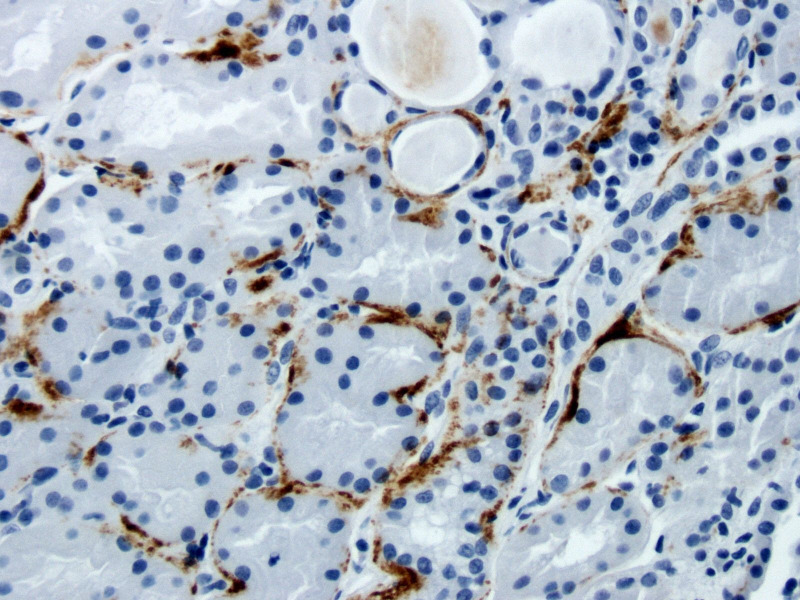
Methods: In this cross-sectional study, LN kidney biopsies were assessed for MAC deposition by staining for Complement C9, a component of the MAC. Chromogenic immunohistochemistry was performed on paraffin-embedded human renal biopsy sections using unconjugated, murine anti-human Complement C9 (Hycult Biotech, clone X197). Tubular C9 staining intensity was analysed as present versus absent. IFTA was defined as minimal (<10%), mild (10%-24%), moderate (25%-50%) and severe (>50%).
Results: Renal biopsies from 30 patients with LN were studied. There were 24 (80%) female sex, mean age (SD) was 33 (12) years old and 23 (77%) had pure/mixed proliferative LN. Tubular C9 staining was present in 7 (23%) biopsies. 27 patients had minimal-to-mild IFTA and 3 patients had moderate IFTA. Among the C9 + patients, 3 (43%) had moderate IFTA as compared with none in the C9- group, p=0.009. C9 + patients had higher median (IQR) proteinuria as compared with C9- patients: 6.2 g (3.3-13.1) vs 2.4 g (1.3-4.6), p=0.001 at the time of biopsy. There was no difference in estimated glomerular filtration rate (eGFR) between the C9 + and C9- groups.
Conclusion: This study demonstrated that tubular MAC deposition is associated with higher degree of IFTA and proteinuria, which are predictors of progression to ESRD. These results suggest that tubular MAC deposition may be useful in classification of LN. Understanding the role of complement in tubulointerstitial injury will also identify new avenues for LN treatment.
Keywords: autoimmunity; inflammation; lupus erythematosus; lupus nephritis; systemic.
© Author(s) (or their employer(s)) 2022. Re-use permitted under CC BY-NC. No commercial re-use. See rights and permissions. Published by BMJ.
Conflict of interest statement
Competing interests: None declared.
Figures
Similar articles
-
Renal Fibrosis in Lupus Nephritis.Int J Mol Sci. 2022 Nov 18;23(22):14317. doi: 10.3390/ijms232214317. Int J Mol Sci. 2022. PMID: 36430794 Free PMC article. Review.
-
Urine Proteomics Link Complement Activation with Interstitial Fibrosis/Tubular Atrophy in Lupus Nephritis Patients.Semin Arthritis Rheum. 2023 Dec;63:152263. doi: 10.1016/j.semarthrit.2023.152263. Epub 2023 Sep 27. Semin Arthritis Rheum. 2023. PMID: 37802003 Free PMC article.
-
Factors associated with worsening interstitial fibrosis/tubular atrophy in lupus nephritis patients undergoing clinically indicated repeat kidney biopsy.BMC Nephrol. 2025 Apr 12;26(1):189. doi: 10.1186/s12882-025-04108-0. BMC Nephrol. 2025. PMID: 40221666 Free PMC article.
-
Predicting kidney outcomes among Latin American patients with lupus nephritis: The prognostic value of interstitial fibrosis and tubular atrophy and tubulointerstitial inflammation.Lupus. 2023 Mar;32(3):411-423. doi: 10.1177/09612033231151597. Epub 2023 Jan 17. Lupus. 2023. PMID: 36647707
-
Deposition of the Membrane Attack Complex in Healthy and Diseased Human Kidneys.Front Immunol. 2021 Feb 11;11:599974. doi: 10.3389/fimmu.2020.599974. eCollection 2020. Front Immunol. 2021. PMID: 33643288 Free PMC article. Review.
Cited by
-
Urinary complement biomarkers in immune-mediated kidney diseases.Front Immunol. 2024 Jun 3;15:1357869. doi: 10.3389/fimmu.2024.1357869. eCollection 2024. Front Immunol. 2024. PMID: 38895123 Free PMC article. Review.
-
Renal Fibrosis in Lupus Nephritis.Int J Mol Sci. 2022 Nov 18;23(22):14317. doi: 10.3390/ijms232214317. Int J Mol Sci. 2022. PMID: 36430794 Free PMC article. Review.
-
Urinary Biomarkers for Lupus Nephritis: A Systems Biology Approach.J Clin Med. 2024 Apr 18;13(8):2339. doi: 10.3390/jcm13082339. J Clin Med. 2024. PMID: 38673612 Free PMC article. Review.
-
Macrophage Infiltration Correlated with IFI16, EGR1 and MX1 Expression in Renal Tubular Epithelial Cells Within Lupus Nephritis-Associated Tubulointerstitial Injury via Bioinformatics Analysis.J Inflamm Res. 2024 Dec 24;17:11469-11483. doi: 10.2147/JIR.S489087. eCollection 2024. J Inflamm Res. 2024. PMID: 39735896 Free PMC article.
-
Microvascular C5b-9 deposition in non-lesional skin in patients with SLE and its correlation with active lupus nephritis: a prospective observational study.Lupus Sci Med. 2023 Oct;10(2):e000996. doi: 10.1136/lupus-2023-000996. Lupus Sci Med. 2023. PMID: 37879755 Free PMC article.
References
-
- Bajema IM, Wilhelmus S, Alpers CE, et al. . Revision of the International Society of Nephrology/Renal pathology Society classification for lupus nephritis: clarification of definitions, and modified National Institutes of health activity and chronicity indices. Kidney Int 2018;93:789–96. 10.1016/j.kint.2017.11.023 - DOI - PubMed
Publication types
MeSH terms
Substances
Grants and funding
LinkOut - more resources
Full Text Sources
Medical
Research Materials
Miscellaneous