. 2022 Jan 11;119(2):e2102953118.
doi: 10.1073/pnas.2102953118.

Lipoprotein-associated phospholipase A2: A paradigm for allosteric regulation by membranes

Affiliations

Lipoprotein-associated phospholipase A2: A paradigm for allosteric regulation by membranes

Varnavas D Mouchlis et al. Proc Natl Acad Sci U S A. .

Abstract

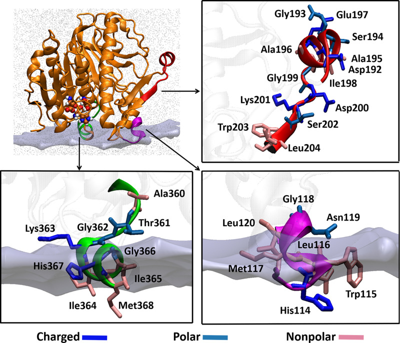
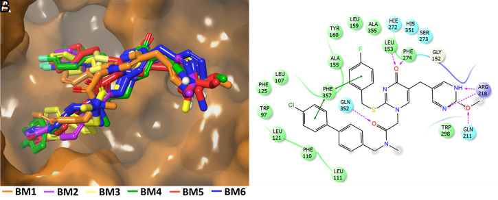
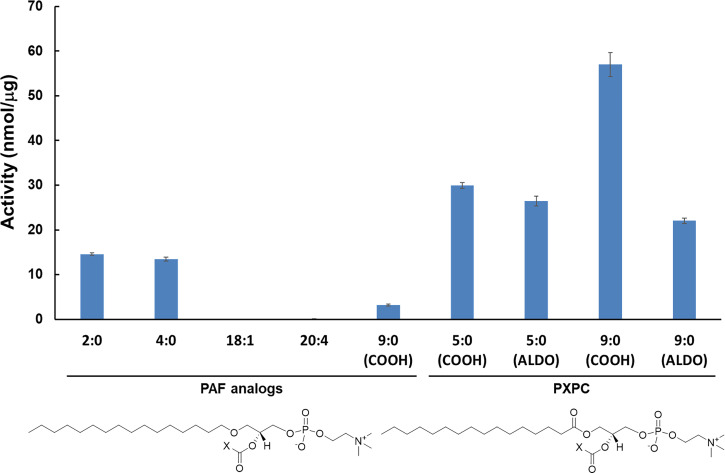
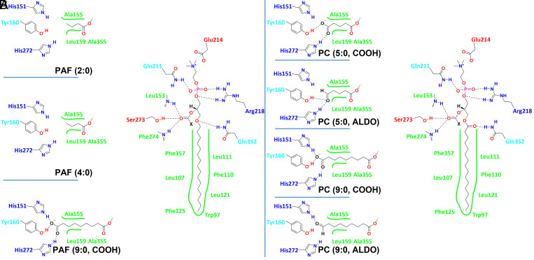
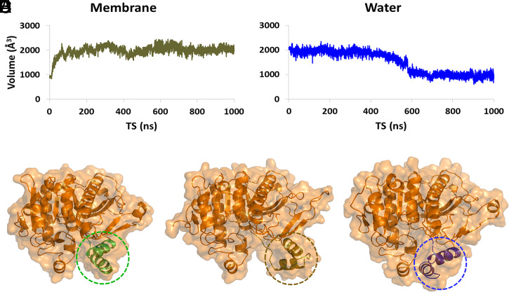
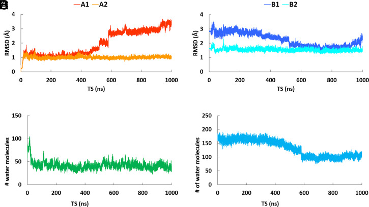
Lipoprotein-associated phospholipase A2 (Lp-PLA2) associates with low- and high-density lipoproteins in human plasma and specifically hydrolyzes circulating oxidized phospholipids involved in oxidative stress. The association of this enzyme with the lipoprotein's phospholipid monolayer to access its substrate is the most crucial first step in its catalytic cycle. The current study demonstrates unequivocally that a significant movement of a major helical peptide region occurs upon membrane binding, resulting in a large conformational change upon Lp-PLA2 binding to a phospholipid surface. This allosteric regulation of an enzyme's activity by a large membrane-like interface inducing a conformational change in the catalytic site defines a unique dimension of allosterism. The mechanism by which this enzyme associates with phospholipid interfaces to select and extract a single phospholipid substrate molecule and carry out catalysis is key to understanding its physiological functioning. A lipidomics platform was employed to determine the precise substrate specificity of human recombinant Lp-PLA2 and mutants. This study uniquely elucidates the association mechanism of this enzyme with membranes and its resulting conformational change as well as the extraction and binding of specific oxidized and short acyl-chain phospholipid substrates. Deuterium exchange mass spectrometry coupled with molecular dynamics simulations was used to define the precise specificity of the subsite for the oxidized fatty acid at the sn-2 position of the phospholipid backbone. Despite the existence of several crystal structures of this enzyme cocrystallized with inhibitors, little was understood about Lp-PLA2's specificity toward oxidized phospholipids.

Keywords: allosterism; lipid; lipoprotein; membrane; phospholipase.


Conflict of interest statement

The authors declare no competing interest.

Figures

Fig. 1.
Fig. 1.
Peptide regions of Lp-PLA2 that interact with phospholipid vesicles and HDL. Residues 114 to 120 and 360 to 368 mediate Lp-PLA2 association with the membrane, while residues 192 to 204 were found to be involved in the association with HDL. These regions were used to generate an in silico enzyme–membrane model.
Fig. 2.
Fig. 2.
Optimized binding mode of SB-402564 after the MD simulations. (A) Each of the six docking binding modes adopted the same conformation and orientation in the Lp-PLA2 active site (Movies S1–S6); see the composite of all six (Movie S7). (B) Noncovalent interactions of the inhibitor with the active site residues are also shown.
Fig. 3.
Fig. 3.
Enzymatic activity of Lp-PLA2 toward 100 µM of various PAF analogs and oxidized phospholipids.
Fig. 4.
Fig. 4.
Binding interactions with the active site residues of Lp-PLA2 revealed by MD simulations for (A) PAF analogs (e.g., PAF-azelaoyl) and (B) oxidized PCs (e.g., azelaoyl-PC and F2-isoprostane-PC).
Fig. 5.
Fig. 5.
Allosteric regulation of Lp-PLA2 by the membrane bilayer (Movie S17). (A) Volume of the Lp-PLA2 active site during the MD simulation in the presence of a membrane patch, (B) volume of the Lp-PLA2 active site during the simulation in water, (C) conformation of Lp-PLA2 as described in the crystal structure (PDB ID: 3D59) used for the MD simulation (circled residues 100 to 130 are colored in green, (D) conformation of Lp-PLA2 after the MD simulation in the presence of a membrane patch (circled residues 100 to 130 are colored in brown), and (E) conformation of Lp-PLA2 after the MD simulation in presence of water (circled residues 100 to 130 are colored in blue).
Fig. 6.
Fig. 6.
RMSD versus time of the simulation for the “open” conformation of Lp-PLA2 in the presence of water. (A, A1) The backbone atoms of Lp-PLA2 in the trajectory were aligned to the starting (“open”) conformation of the simulation (red color), (A, A2) same alignment as A1, but the peptide region 100 to 130 was excluded (orange color), (B, B1) The backbone atoms of Lp-PLA2 in the trajectory were aligned to the crystallographic conformation (PDB ID: 3D59, blue color), (B, B2) same alignment as B1, but the peptide region 100 to 130 was excluded (cyan color). (C) Number of water molecules in the active site of Lp-PLA2 versus time of the simulation in the presence of a membrane patch, and (D) number of water molecules in the active site of Lp-PLA2 versus time of the simulation in the presence of water.

References

    1. Dennis E. A., Cao J., Hsu Y.-H., Magrioti V., Kokotos G., Phospholipase A2 enzymes: Physical structure, biological function, disease implication, chemical inhibition, and therapeutic intervention. Chem. Rev. 111, 6130–6185 (2011). - PMC - PubMed
    1. Mouchlis V. D., Bucher D., McCammon J. A., Dennis E. A., Membranes serve as allosteric activators of phospholipase A2, enabling it to extract, bind, and hydrolyze phospholipid substrates. Proc. Natl. Acad. Sci. U.S.A. 112, E516–E525 (2015). - PMC - PubMed
    1. Cao J., Burke J. E., Dennis E. A., Using hydrogen/deuterium exchange mass spectrometry to define the specific interactions of the phospholipase A2 superfamily with lipid substrates, inhibitors, and membranes. J. Biol. Chem. 288, 1806–1813 (2013). - PMC - PubMed
    1. Stafforini D. M., Elstad M. R., McIntyre T. M., Zimmerman G. A., Prescott S. M., Human macrophages secret platelet-activating factor acetylhydrolase. J. Biol. Chem. 265, 9682–9687 (1990). - PubMed
    1. McIntyre T. M., Prescott S. M., Stafforini D. M., The emerging roles of PAF acetylhydrolase. J. Lipid Res. 50 (suppl.), S255–S259 (2009). - PMC - PubMed

Publication types