

Single cell analysis reveals inhibition of angiogenesis attenuates the progression of heterotopic ossification in Mkx-/- mice
- PMID: 34996891
- PMCID: PMC8741758
- DOI: 10.1038/s41413-021-00175-9
Single cell analysis reveals inhibition of angiogenesis attenuates the progression of heterotopic ossification in Mkx-/- mice
Abstract
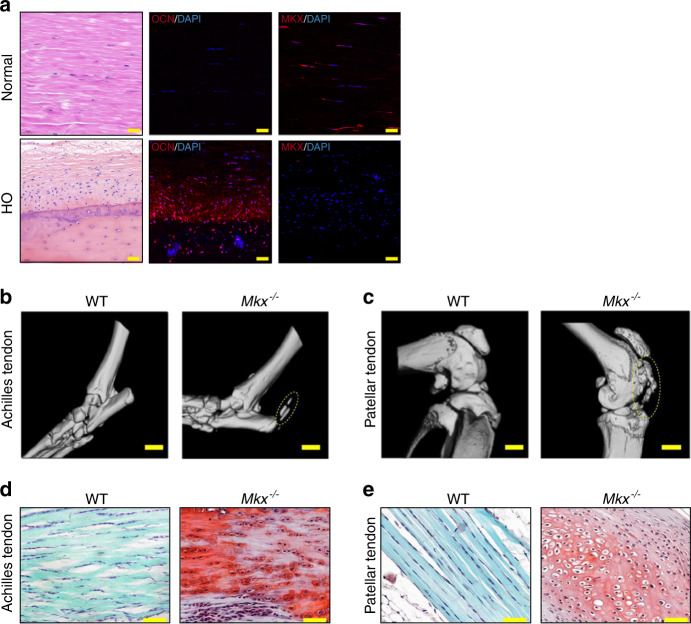
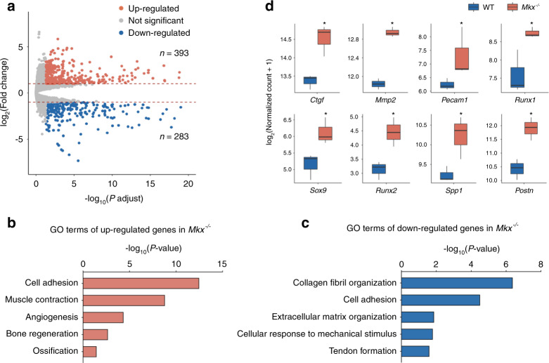
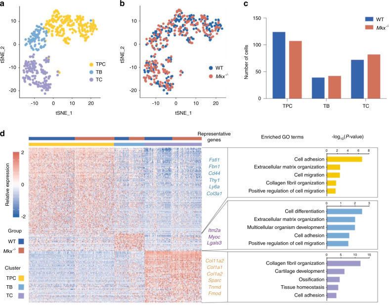
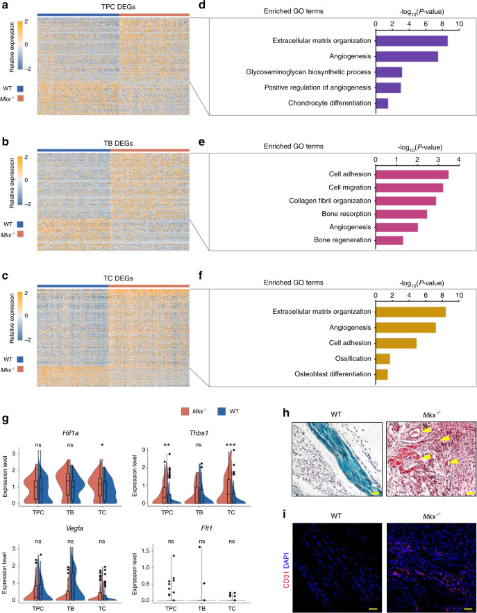
Tendon heterotopic ossification (HO) is characterized by bone formation inside tendon tissue, which severely debilitates people in their daily life. Current therapies fail to promote functional tissue repair largely due to our limited understanding of HO pathogenesis. Here, we investigate the pathological mechanism and propose a potential treatment method for HO. Immunofluorescence assays showed that the Mohawk (MKX) expression level was decreased in human tendon HO tissue, coinciding with spontaneous HO and the upregulated expression of osteochondrogenic and angiogenic genes in the tendons of Mkx-/- mice. Single-cell RNA sequencing analyses of wild-type and Mkx-/- tendons identified three cell types and revealed the excessive activation of osteochondrogenic genes during the tenogenesis of Mkx-/- tendon cells. Single-cell analysis revealed that the gene expression program of angiogenesis, which is strongly associated with bone formation, was activated in all cell types during HO. Moreover, inhibition of angiogenesis by the small-molecule inhibitor BIBF1120 attenuated bone formation and angiogenesis in the Achilles tendons of both Mkx mutant mice and a rat traumatic model of HO. These findings provide new insights into the cellular mechanisms of tendon HO and highlight the inhibition of angiogenesis with BIBF1120 as a potential treatment strategy for HO.
© 2022. The Author(s).
Conflict of interest statement
The authors declare no competing interests.
Figures


Similar articles
-
Gene targeting of the transcription factor Mohawk in rats causes heterotopic ossification of Achilles tendon via failed tenogenesis.Proc Natl Acad Sci U S A. 2016 Jul 12;113(28):7840-5. doi: 10.1073/pnas.1522054113. Epub 2016 Jul 1. Proc Natl Acad Sci U S A. 2016. PMID: 27370800 Free PMC article.
-
Mkx-Deficient Mice Exhibit Hedgehog Signaling-Dependent Ectopic Ossification in the Achilles Tendons.J Bone Miner Res. 2019 Mar;34(3):557-569. doi: 10.1002/jbmr.3630. Epub 2019 Feb 25. J Bone Miner Res. 2019. PMID: 30458056 Free PMC article.
-
Suppression of TNF-α activity by immobilization rescues Mkx expression and attenuates tendon ossification in a mouse Achilles tenotomy model.J Orthop Res. 2024 Oct;42(10):2140-2148. doi: 10.1002/jor.25906. Epub 2024 May 28. J Orthop Res. 2024. PMID: 38806292
-
Heterotopic ossification of tendon and ligament.J Cell Mol Med. 2020 May;24(10):5428-5437. doi: 10.1111/jcmm.15240. Epub 2020 Apr 15. J Cell Mol Med. 2020. PMID: 32293797 Free PMC article. Review.
-
Challenges of heterotopic ossification-Molecular background and current treatment strategies.Clin Exp Pharmacol Physiol. 2018 Dec;45(12):1229-1235. doi: 10.1111/1440-1681.13025. Epub 2018 Oct 4. Clin Exp Pharmacol Physiol. 2018. PMID: 30144316 Review.
Cited by
-
Single-cell RNA sequencing in orthopedic research.Bone Res. 2023 Feb 24;11(1):10. doi: 10.1038/s41413-023-00245-0. Bone Res. 2023. PMID: 36828839 Free PMC article. Review.
-
Macrophage-Derived TGF-β and VEGF Promote the Progression of Trauma-Induced Heterotopic Ossification.Inflammation. 2023 Feb;46(1):202-216. doi: 10.1007/s10753-022-01723-z. Epub 2022 Aug 20. Inflammation. 2023. PMID: 35986177
-
Intersections of Fibrodysplasia Ossificans Progressiva and Traumatic Heterotopic Ossification.Biomolecules. 2024 Mar 14;14(3):349. doi: 10.3390/biom14030349. Biomolecules. 2024. PMID: 38540768 Free PMC article. Review.
-
Single-cell and spatial transcriptomics reveal changes in cell heterogeneity during progression of human tendinopathy.BMC Biol. 2023 Jun 6;21(1):132. doi: 10.1186/s12915-023-01613-2. BMC Biol. 2023. PMID: 37280595 Free PMC article.
-
Development of an Animal Model for Traumatic Brain Injury Augmentation of Heterotopic Ossification in Response to Local Injury.Biomedicines. 2023 Mar 18;11(3):943. doi: 10.3390/biomedicines11030943. Biomedicines. 2023. PMID: 36979922 Free PMC article.
References
-
- Shehab D, Elgazzar AH, Collier BD. Heterotopic ossification. J. Nucl. Med. 2002;43:346–353. - PubMed
Grants and funding
- 2017YFA0104902/Ministry of Science and Technology of the People's Republic of China (Chinese Ministry of Science and Technology)
- 81330041/National Natural Science Foundation of China (National Science Foundation of China)
- 81522029/National Natural Science Foundation of China (National Science Foundation of China)
- 2013TD11/Science and Technology Department of Zhejiang Province
LinkOut - more resources
Full Text Sources
Molecular Biology Databases
