Observational Study
. 2022 Jan 7;12(1):88.
doi: 10.1038/s41598-021-03716-8.

Baseline predictors for subretinal fibrosis in neovascular age-related macular degeneration

Affiliations
Observational Study

Baseline predictors for subretinal fibrosis in neovascular age-related macular degeneration

Philipp K Roberts et al. Sci Rep. .

Abstract

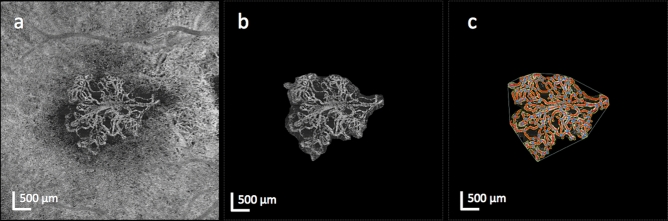
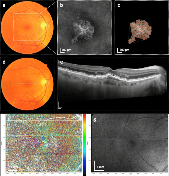
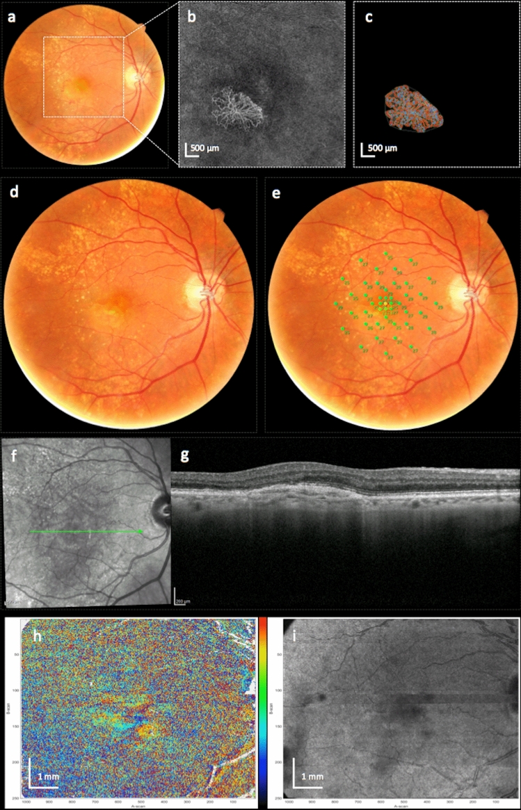
To find baseline predictors for subretinal fibrosis (SF) in neovascular age-related macular degeneration (nAMD). Forty-five eyes of 45 participants with treatment-naïve nAMD were consecutively enrolled and treated according to a standardized treat-and-extend protocol. Spectral-domain optical coherence tomography (OCT), color fundus photography and fluorescein angiography as well as novel imaging modalities polarization-sensitive OCT and OCT angiography (OCTA) were performed to detect SF after 1 year and find baseline predictors for SF development. Baseline OCTA scans were evaluated for quantitative features such as lesion area, vessel area, vessel junctions, vessel length, vessel endpoints and mean lacunarity. Additionally, the type of macular neovascularization, the presence of subretinal fluid, intraretinal fluid (IRF), subretinal hyperreflective material (SHRM), retinal hemorrhage as well as best-corrected visual acuity (BCVA) were evaluated. After 12 months 8 eyes (18%) developed SF. Eyes with SF had worse baseline BCVA (p = .001) and a higher prevalence of IRF (p = .014) and SHRM at baseline (p = .017). There was no significant difference in any of the evaluated quantitative OCTA parameters (p > .05) between eyes with and without SF. There were no quantitative baseline microvascular predictors for SF in our study. Low baseline BCVA, the presence of IRF and SHRM, however, are easily identifiable baseline parameters indicating increased risk.


Conflict of interest statement

The authors declare no competing interests.

Figures

Figure 1
Figure 1
Representative swept-source optical coherence tomography angiography (OCTA) en face imaging of a left eye with a type 1 macular neovascularization (MNV) (a). After careful manual segmentation of the MNV lesion (b) in FIJI, the Angiotool program was used for quantitative analysis of the microvascular complex. The result of the analysis (c) shows the skeletonized vessels (red lines), junctions (blue dots), lesion area (outer white line) and endpoints (points of open-ended vessels).
Figure 2
Figure 2
Left eye of a patient with a mixed type MNV, who developed subretinal fibrosis (SF). Figures show the MNV at baseline (ac) and after 12 months (dg). Color fundus photography at baseline (a) and after 12 months (d) shows the development of yellow subretinal material, colocalized with an area of subretinal hyperreflective material in the SD-optical coherence tomography (OCT) B-scan at month 12 (e). Axis orientation (f) and pseudo-SLO PS-OCT imaging (g) with automatically segmented fibrosis (highlighted in blue) illustrate the area of the subretinal fibrosis. Color bar indicates − 90 to + 90° for axis orientation. Baseline en face OCT angiography (b) shows a subfoveal circular neovascular net. The result of the quantitative vessel analysis using the Angiotool software is shown in (c). The location of the OCTA en face images is indicated by the white square in (a) and the location of the SD-OCT B-scan is indicated by the white dotted line in (d).
Figure 3
Figure 3
Right eye of a patient with a type 1 MNV, who did not develop subretinal fibrosis. Figures show the MNV lesion at baseline (ac) and after 12 months (dg). Color fundus photography at baseline (a) and after 12 months (d) show no presence of subretinal whitish-yellow material. SD-optical coherence tomography (OCT) at month 12 (e) shows a fibrovascular pigment epithelial detachment but no signs of exudation such as subretinal fluid or intraretinal fluid. Axis orientation (f) and pseudo-SLO PS-OCT imaging (g) show no presence of fibrosis. Color bar indicates − 90 to + 90° for axis orientation Baseline en face OCT angiography (b) as well as the result of the quantitative vessel analysis (c) using the Angiotool software is shown. The location of the OCTA en face images is indicated by the white square in (a) and the location of the SD-OCT B-scan is indicated by the white dotted line in (d).
Figure 4
Figure 4
Right eye of a patient with a type 2 MNV and questionable development of subretinal fibrosis. Figures show the MNV lesion at baseline (ac) and after 12 months (di). The patient had a small hemorrhage on color fundus photography at baseline (a) and developed questionable subretinal fibrosis (SF) after 12 months visible as faint yellowish subretinal material in (d). The area is colocalized with a fibrovascular pigment epithelial detachment in the SD-optical coherence tomography (OCT) B-scan at month 12 (g). The location of the B-scan is indicated by the green arrow in the infrared en face image (f). The baseline en face OCT angiography image (b) and the quantitative vessel analysis (c) using the Angiotool software is shown and the location of the OCT angiography en face images is indicated by the white dotted square in (a). Microperimetry (e) shows well-preserved retinal function indicated by green dots in the area with presumed fibrosis. Axis orientation (h) and pseudo-SLO PS-OCT imaging (i) do not confirm the presence of fibrosis. Using multimodal imaging the lesion was therefore graded as no fibrosis. Color bar indicates − 90 to + 90° for axis orientation.

Similar articles

Cited by

References

    1. Cheung CMG, et al. The evolution of fibrosis and atrophy and their relationship with visual outcomes in asian persons with neovascular age-related macular degeneration. Ophthalmol. Retina. 2019;3:1045–1055. doi: 10.1016/j.oret.2019.06.002. - DOI - PubMed
    1. Wolff B, et al. Ten-year outcomes of anti-vascular endothelial growth factor treatment for neovascular age-related macular disease: A single-centre French study. Clin. Exp. Ophthalmol. 2020;48:636–643. doi: 10.1111/ceo.13742. - DOI - PubMed
    1. Hogg R, et al. Identification of lesion components that influence visual function in age related macular degeneration. Br. J. Ophthalmol. 2003;87:609–614. doi: 10.1136/bjo.87.5.609. - DOI - PMC - PubMed
    1. Gillies M, et al. Ten-year treatment outcomes of neovascular age-related macular degeneration from two regions. Am. J. Ophthalmol. 2020;210:116–124. doi: 10.1016/j.ajo.2019.10.007. - DOI - PubMed
    1. Daniel E, et al. Development and course of scars in the comparison of age-related macular degeneration treatments trials. Ophthalmology. 2018;125:1037–1046. doi: 10.1016/j.ophtha.2018.01.004. - DOI - PMC - PubMed

Publication types

MeSH terms

Substances