. 2022 Jan 7;12(1):8.
doi: 10.1038/s41598-021-03795-7.

Decrease of Pdzrn3 is required for heart maturation and protects against heart failure

Affiliations

Decrease of Pdzrn3 is required for heart maturation and protects against heart failure

Mathieu Pernot et al. Sci Rep. .

Abstract

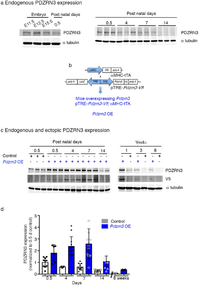
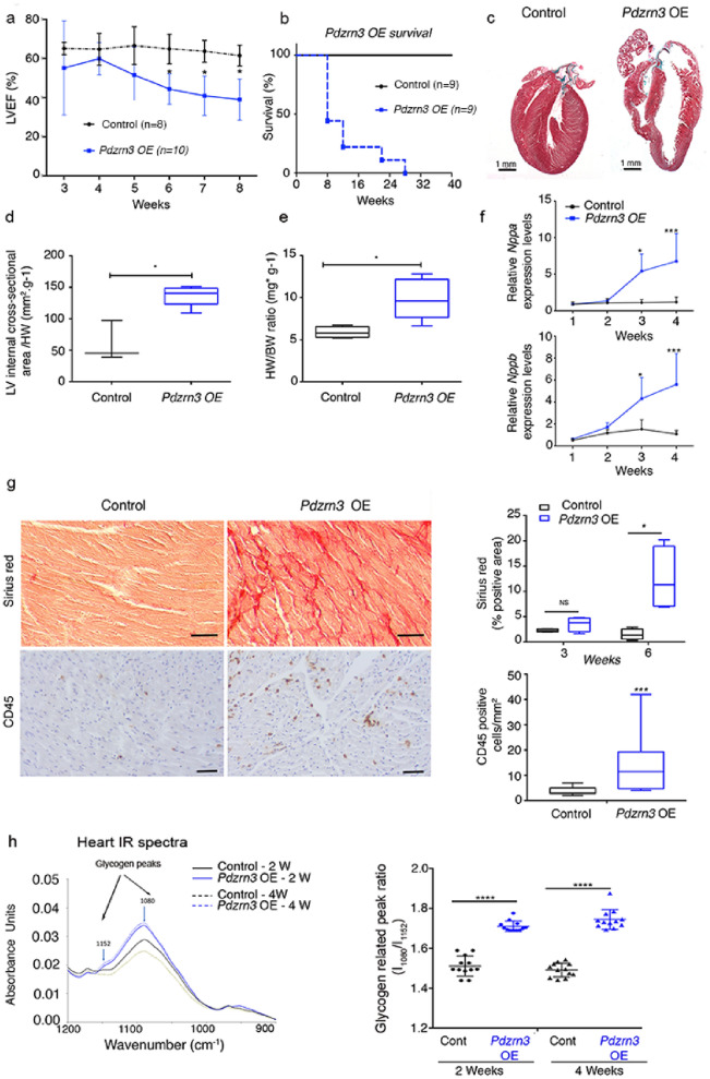
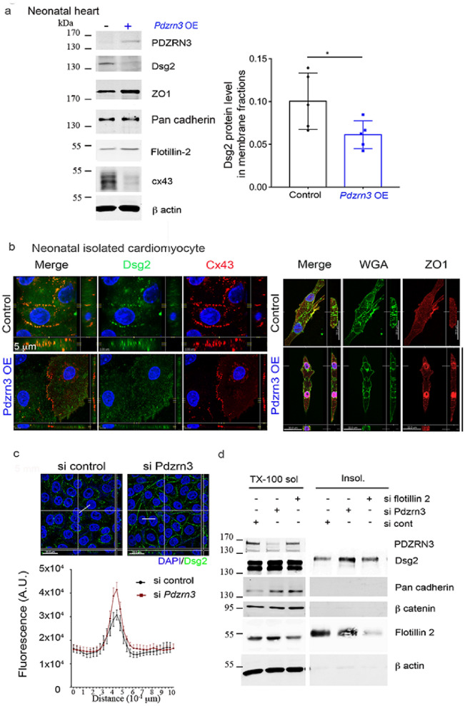
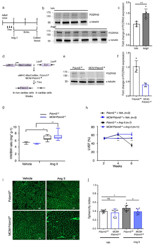
Heart failure is the final common stage of most cardiopathies. Cardiomyocytes (CM) connect with others via their extremities by intercalated disk protein complexes. This planar and directional organization of myocytes is crucial for mechanical coupling and anisotropic conduction of the electric signal in the heart. One of the hallmarks of heart failure is alterations in the contact sites between CM. Yet no factor on its own is known to coordinate CM polarized organization. We have previously shown that PDZRN3, an ubiquitine ligase E3 expressed in various tissues including the heart, mediates a branch of the Planar cell polarity (PCP) signaling involved in tissue patterning, instructing cell polarity and cell polar organization within a tissue. PDZRN3 is expressed in the embryonic mouse heart then its expression dropped significantly postnatally corresponding with heart maturation and CM polarized elongation. A moderate CM overexpression of Pdzrn3 (Pdzrn3 OE) during the first week of life, induced a severe eccentric hypertrophic phenotype with heart failure. In models of pressure-overload stress heart failure, CM-specific Pdzrn3 knockout showed complete protection against degradation of heart function. We reported that Pdzrn3 signaling induced PKC ζ expression, c-Jun nuclear translocation and a reduced nuclear ß catenin level, consistent markers of the planar non-canonical Wnt signaling in CM. We then show that subcellular localization (intercalated disk) of junction proteins as Cx43, ZO1 and Desmoglein 2 was altered in Pdzrn3 OE mice, which provides a molecular explanation for impaired CM polarization in these mice. Our results reveal a novel signaling pathway that controls a genetic program essential for heart maturation and maintenance of overall geometry, as well as the contractile function of CM, and implicates PDZRN3 as a potential therapeutic target for the prevention of human heart failure.


Conflict of interest statement

The authors declare no competing interests.

Figures

Figure 1
Figure 1
Physiological PDZRN3 expression around birth. Results of Pdzrn3 overexpression. (a) Representative Western blot demonstrates levels of PDZRN3 in the embryo at E11.5, 12.5 and 15.5 days; and after birth (0.5, 4, 7 and 14 days). αtubulin was used as the loading control. (b) Schematic of Pdzrn3 overexpression (OE) in the heart. Control mice were (MHC-tTA, Pdzrn3 OE mice were pTRE-Pdzrn3; αMHC-tTA. (c) Representative Western blot demonstrates levels of endogenous PDZRN as well as overexpression from 0.5 to 14 days and at 8 weeks after birth. αtubulin was used as the loading control. (d) The ratio is quantified and calibrated to the average of 0.5 d control mice. Data are expressed as mean ± s.e.m. (n = 6–13).
Figure 2
Figure 2
Myocardial reactivation of Pdzrn3 induces eccentric cardiac hypertrophy. (a) LVEF was quantified from 3 to 8 weeks of age (control, n = 4; Pdzrn3 OE, n = 7). (b) Kaplan–Meier curve showing survival rate of control and Pdzrn3 OE mice from birth until 40 weeks of age (control and Pdzrn3 OE groups, n = 9). (c) Representative images of hematoxylin and eosin (H&E) stained sections of control and Pdzrn3 OE hearts at 6 weeks, demonstrate that Pdzrn3 OE mice have enlarged hearts. (d) Increased LV internal cross section (control, n = 3; Pdzrn3 OE, n = 5). (e) Increased heart weight (HW) to body weight (BW) ratio (control, n = 5; Pdzrn3 OE, n = 5). (f) Increase of the mRNA levels of atrial (Nppa) and brain (Nppb) natriuretic peptide in Pdzrn3 OE versus control mice over time. mRNA levels were normalized to cyclophiline and are expressed as a relative expression/fold increase over levels found in control mice at 1 week (n = 3–7 mice per group). (g) In hearts from Pdzrn3 OE mice, quantification of total collagen deposition on picrosirius red staining revealing an increase in fibrosis and CD45 immunolabeling, and an increase of inflammatory cells at 6 and 4 weeks of age, respectively (n = 4 mice per group) (the scale bars represent 50 μm). (h) FTIR spectra after pre-processing and classification (n = 6 spectra per condition) in the 900 cm−1 to 1200 cm−1 spectral region obtained heart tissue cryo-sections retrieved from the left ventricle from control and Pdzrn3 OE mice (n = 3 per group at 2 and 4 weeks). Arrows indicate the wavenumbers studied. Changes in the heart glycogen illustrated with the absorbance ratio 1080 cm−1/1152 cm−1. All the data are represented as mean ± sem, with n = 3 mice. Mean ± s.e.m. *P < 0.05, **P < 0.005, ***P < 0.001 by repeated-measures two way ANOVA with post-hoc sidak’s test (a, f); Kaplan–Meier nonparametric regression analysis and the log-rank test (b) and unpaired t-test (d, e, g).
Figure 3
Figure 3
Cardiac specific overexpression of Pdzrn3 impairs cardiomyocyte elongation. (a) The sphericity index was determined by measuring the area (A) and the circumference (P) of myocytes. The ratio of an “ideal” round cell is close to 1, whereas that of an elongated cell is closer to 0. Cardiomyocytes that were in the longitudinal plane were selected for quantification of the sphericity index. (b) Representative images of control and Pdzrn3 OE heart sections stained with wheat germ agglutinin (WGA)-FITC (scale bars represent 20 μm) at 3 W. Quantification of myocyte cross-sectional sphericity of control and Pdzrn3 OE heart sections. Data are represented as mean ± sem. Significance vs control **P < 0.01 by one way ANOVA plus bonferroni test (control vs. Pdzrn3 OE group, n = 4 at 2 weeks, n = 3 at 3 weeks and n = 5 at 4 weeks). (c) Representative electron micrographs showing morphological disorganization of cardiomyocytes of control and Pdzrn3 OE heart at 8 weeks of age (scale bars represent 1 μm). (d) Quantitative F-actin/G-actin ratios in heart lysates from 2-week-old mice were measured with actin polymerization in vivo assay kit. Western blot analysis of G-actin (G) and F-actin (F) fraction from WT and Pdzrn3 OE heart extracts were probed with anti-actin antibody. Ratios of F-actin/G actin was determined from the blots by optical density measurements. **P < 0.001, by unpaired t-test. (e) Western blot analysis of indicated proteins in heart tissues retrieved from control and Pdzrn3 OE mice at 2 weeks (W) of age. Relative expression of Pkc ζand c-Jun from Western blots. Significance vs. control **P < 0.01; ***P < 0.001 by unpaired t-test (at 1 week, n = 4 mice per group; at 2 weeks, n = 6 mice per group). (f) Western blot analysis of indicated proteins in heart tissue retrieved from control and Pdzrn3 OE mice after tissue fractionation to isolate cytosolic, membrane and nuclear (soluble) fractions. (n = 3 mice per group).
Figure 4
Figure 4
Myocardial reactivation of Pdzrn3 alters postnatal cardiomyocyte maturation. (a) Electron micrographs of part of an intercalated disk between two cardiac of control and Pdzrn3 OE heart at 8 weeks of age (scale bars represent 2 μm) and in right part, schema of ID distribution. (b) Immunolabeling of Cx43, ZO1, β catenin and α actinin in hearts from control and Pdzrn3 OE mice at 2 weeks. Scale bars represent 10 μm. (c) Western blot analysis of indicated proteins in heart tissues retrieved from control and Pdzrn3 OE mice at 1 and 2 weeks (W) of age. Relative expression of Cx43, from Western blots. Significance vs. control *P < 0.05; ***P < 0.001 by unpaired t-test (at 1 week, n = 4 mice per group; at 2 weeks, n = 6 mice per group).
Figure 5
Figure 5
Altered subcellular distribution of junction proteins in cell–cell membrane under Pdzrn3 induced signaling. (a) Western blot analysis of indicated proteins in plasma membrane fraction only of heart tissues retrieved from control and Pdzrn3 OE mice at 2 Weeks of age. Relative expression of Dsg2, from Western blots. Significance vs. control *P < 0.05; 1 by unpaired t-test (n = 5 mice per group). (b) Representative confocal microscopy images of primary cardiomyocytes from control and Pdzrn3 OE neonates stained with antibodies against Dsg2, Cx43, WGA-Fitc and ZO1. (c) After immunostaining of HeLa cells depleted of Pdzrn3 (si Pdzrn3) or not (si control) for Dsg2 (green), fluorescence intensity profile curve were plotted. Data are represented as mean ± sem from three independent experiments. (d) Western blot analysis of indicated proteins in triton-X-100 soluble and insoluble fractions from HeLa depleted either of Pdzrn3 (si Pdzrn3) or flotillin-2 (si flotillin 2).
Figure 6
Figure 6
Postnatal time window for cardiac cell differentiation. (a, b) Schematic study timeline of doxycline treatment administration (Dox), tissue collection and echocardiography follow up. (c, e) Immunolabeling of Cx43 and ZO1 in hearts from control and Pdzrn3 OE mice following doxycycline treatment. Scale bars represent 50 μm. (d, f) LVEF quantification under doxycycline treatment in control and Pdzrn3 OE mice in d protocol, respectively n = 6 vs. 8; in h protocol, respectively n = 6 vs. 4 mice.
Figure 7
Figure 7
Depletion of Pdzrn3 protects against decompensated cardiac hypertrophy. (a) Schematic study plan showing timeline of tamoxifen administration (Tmx), Ang II pump implantation, echocardiography (Echo.) follow up and tissue collection. (b) Western blot reporting levels of PDZRN3 in the adult mice under sham condition (Veh) and after AngII treatment. αtubulin was used as the loading control. (c) PDZRN3 ratio is quantified in veh and AngII groups and calibrated to the average of veh mice. Data are expressed as mean ± SD. Significance vs. control **P < 0.001 by unpaired t-test (n = 4 in veh- and n = 6 in AngII treated-groups). (d) Schematic of Pdzrn3 deletion in cardiac cells. Control mice were Pdzrn3f/f. MCM-Pdzrn3 KO mice were MHC-MerCreMer; Pdzrn3f/f. (e) Representative Western blot demonstrates conditional depletion of Pdzrn3 in MCM-Pdzrn3f/f mice compared to Pdzrn3f/f control mice. αtubulin was used as the loading control. (f) The ratio is quantified and calibrated to the average of Pdzrn3f/f control mice. Data are expressed as mean ± s.d. Significance vs. control *P < 0.05 by unpaired t-test (n = 3 per group). (g) Ratio of heart weight (HW) to body weight (BW). (h) Quantification of left ventricular ejection fraction (LVEF) in tamoxifen treated Pdzrn3f/f and MCM-Pdzrn3 mice following Ang II treatment (vehicle treated groups, n = 3; Ang II-treated groups, Pdzrn3f/f n = 7, MCM-Pdzrn3, n = 11). (i) Representative images of heart sections from f Pdzrn3f/f and MCM-Pdzrn3f/f mice following either Ang II or Veh treatment, stained with wheat germ agglutinin (WGA)-FITC (scale bars represent 20 μm). (j) Quantification of the cardiomyocyte sphericity index in tamoxifen treated Pdzrn3f/f and MCM-Pdzrn3f/f mice following either Ang II or Veh treatment. (n = 9 per group). *P < 0.05, **P < 0.01. One-way ANOVA with post-hoc tukey’s test.

References

    1. Balse E, Steele DF, Abriel H, Coulombe A, Fedida D, Hatem SN. Dynamic of ion channel expression at the plasma membrane of cardiomyocytes. Physiol. Rev. 2012;92:1317–1358. - PubMed
    1. Le Garrec J-F, Ragni CV, Pop S, Dufour A, Olivo-Marin J-C, Buckingham ME, Meilhac SM. Quantitative analysis of polarity in 3D reveals local cell coordination in the embryonic mouse heart. Development. 2013;140:395–404. - PubMed
    1. Yang Y, Mlodzik M. Wnt-frizzled/planar cell polarity signaling: Cellular orientation by facing the wind (Wnt) Annu. Rev. Cell Dev. Biol. 2015;31:623–646. - PMC - PubMed
    1. Pandur P, Lasche M, Eisenberg LM, Kuhl M. Wnt-11 activation of a non-canonical Wnt signalling pathway is required for cardiogenesis. Nature. 2002;418:636–641. - PubMed
    1. Nagy I, Railo A, Rapila R, Hast T, Sormunen R, Tavi P, Rasanen J, Vainio SJ. Wnt-11 signalling controls ventricular myocardium development by patterning N-cadherin and beta-catenin expression. Cardiovasc. Res. 2010;85:100–109. - PubMed

Publication types