. 2022 Jan 7;12(1):53.
doi: 10.1038/s41598-021-03990-6.

In silico construction of a multiepitope Zika virus vaccine using immunoinformatics tools

Affiliations

In silico construction of a multiepitope Zika virus vaccine using immunoinformatics tools

Ana Clara Barbosa Antonelli et al. Sci Rep. .

Abstract

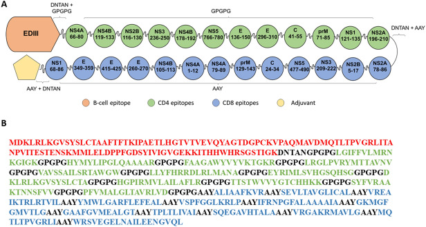
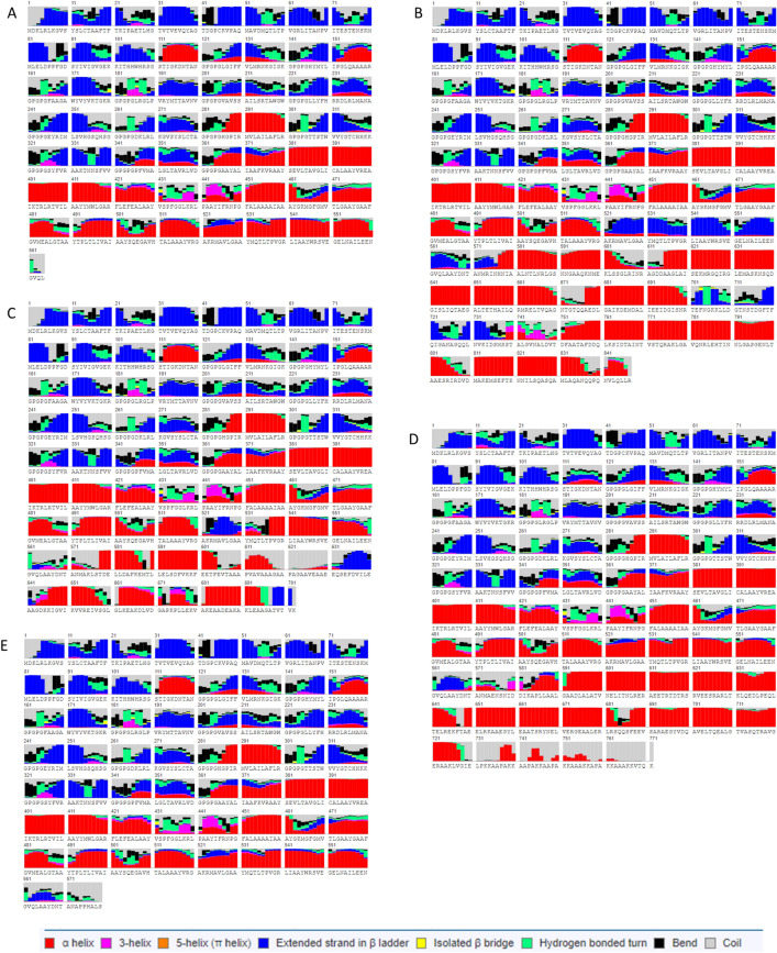
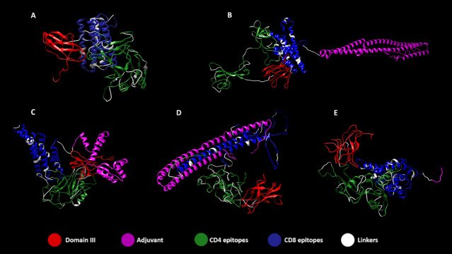
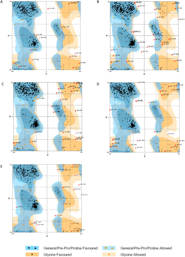
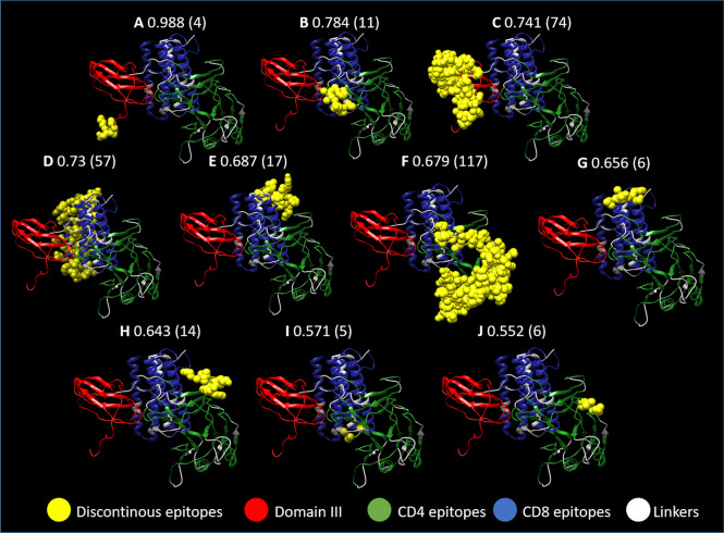
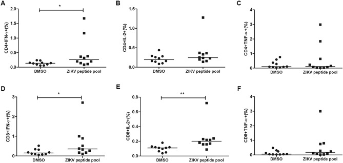
Zika virus (ZIKV) is an arbovirus from the Flaviviridae family and Flavivirus genus. Neurological events have been associated with ZIKV-infected individuals, such as Guillain-Barré syndrome, an autoimmune acute neuropathy that causes nerve demyelination and can induce paralysis. With the increase of ZIKV infection incidence in 2015, malformation and microcephaly cases in newborns have grown considerably, which suggested congenital transmission. Therefore, the development of an effective vaccine against ZIKV became an urgent need. Live attenuated vaccines present some theoretical risks for administration in pregnant women. Thus, we developed an in silico multiepitope vaccine against ZIKV. All structural and non-structural proteins were investigated using immunoinformatics tools designed for the prediction of CD4 + and CD8 + T cell epitopes. We selected 13 CD8 + and 12 CD4 + T cell epitopes considering parameters such as binding affinity to HLA class I and II molecules, promiscuity based on the number of different HLA alleles that bind to the epitopes, and immunogenicity. ZIKV Envelope protein domain III (EDIII) was added to the vaccine construct, creating a hybrid protein domain-multiepitope vaccine. Three high scoring continuous and two discontinuous B cell epitopes were found in EDIII. Aiming to increase the candidate vaccine antigenicity even further, we tested secondary and tertiary structures and physicochemical parameters of the vaccine conjugated to four different protein adjuvants: flagellin, 50S ribosomal protein L7/L12, heparin-binding hemagglutinin, or RS09 synthetic peptide. The addition of the flagellin adjuvant increased the vaccine's predicted antigenicity. In silico predictions revealed that the protein is a probable antigen, non-allergenic and predicted to be stable. The vaccine's average population coverage is estimated to be 87.86%, which indicates it can be administered worldwide. Peripheral Blood Mononuclear Cells (PBMC) of individuals with previous ZIKV infection were tested for cytokine production in response to the pool of CD4 and CD8 ZIKV peptide selected. CD4 + and CD8 + T cells showed significant production of IFN-γ upon stimulation and IL-2 production was also detected by CD8 + T cells, which indicated the potential of our peptides to be recognized by specific T cells and induce immune response. In conclusion, we developed an in silico universal vaccine predicted to induce broad and high-coverage cellular and humoral immune responses against ZIKV, which can be a good candidate for posterior in vivo validation.


Conflict of interest statement

The authors declare no competing interests.

Figures

Figure 1
Figure 1
Phylogenetic tree of the 40 complete ZIKV strains used for obtaining the consensus sequence (A). (B) Polyprotein of the ZIKV from the Brazilian consensus sequence. The ZIKV genome codes for seven non-structural proteins (NS1, NS2A, NS2B, NS3, NS4A, NS4B and NS5) and four structural proteins (Capsid protein C, Protein prM, ER anchor for capsid protein C and Envelope protein E). Protein prM is further digested into Peptide pr and Small envelope protein M. ER anchor for capsid protein C is removed by serine protease NS3. The consensus sequence was obtained from 40 complete Brazilian sequences deposited on NCBI.
Figure 2
Figure 2
ZIKVac structure showing the position of CD4 (green) and CD8 epitopes (blue) envelope protein E Domain III (EDIII) and adjuvant (yellow pentagon) (A). The protein sequence of and ZIKVac sequence without conjugated adjuvants showing the EDIII (red), CD4 epitopes (green), CD8 epitopes (blue) and linkers (black) (B). DNTAN linkers were used to separate EDIII and adjuvant from the epitopes, GPGPG linkers were used between CD4 epitopes and AAY linkers between CD8 epitopes.
Figure 3
Figure 3
Secondary structure of the vaccine protein alone (A) and in association with adjuvants (B–E). Helices, coils and beta-sheets are specified in the legend, to note: alpha helix in red, 3-helix in magenta, 5-helix (pi helix) in orange, extended strand in beta ladder in blue, isolated beta bridge in yellow, hydrogen bonded turn in green, bend in black and coil in gray. ZIKVac + Flagellin (B), 50S ribosomal (C), HBH (D), RS09 (E).
Figure 4
Figure 4
Tertiary structure of ZIKVac (A) with highlighted epitopes flanked by linkers and the adjuvants at the C-terminal position, which are the flagellin (B), 50S Ribossomal (C), Heparin-binding hemagglutinin (HBH) (D) and RS09 (E). In red is the ZIKV Envelope Domain III, in magenta the adjuvants, in green CD4 epitopes, in blue CD8 epitopes and in white the linkers.
Figure 5
Figure 5
Ramachandran plots of the ZIKVac alone (A) and linked with the adjuvants, which are the flagellin (B), 50S Ribosomal protein (C), Heparin-binding hemagglutinin (HBH) (D) and RS09 (E). Black squares and triangles in blue areas indicate General/Pre-Pro/Proline Favored, orange squares and triangles in lighter blue areas indicate General/Pre-Pro/Proline Allowed, black Xs in orange regions indicate Glycine Favored and Orange Xs in lighter orange regions indicate Glycine allowed.
Figure 6
Figure 6
Discontinuous epitopes present in the vaccine construct. Two epitopes are located at the Envelope Domain III portion of the protein (A and C) with some of the highest scores (0.988 and 0.741) and with 4 and 74 amino acids, respectively. The other discontinuous epitopes are distributed among the CD4 and CD8 epitopes (B, DJ). In yellow are the predicted discontinuous epitopes, in red the domain III from the ZIKV Envelope protein, in green CD4 epitopes, in blue CD8 epitopes and in white the linkers.
Figure 7
Figure 7
CD4 + and CD8 + T cell responses to ZIKV peptides. PBMC of 10 individuals with previous infection to ZIKV were stimulated for 5 h with the ZIKV peptide pool. After 1 h of stimulation, Brefeldin A was added. The cells were stained with antibodies for the surface markers CD3, CD4 and CD8 and intracellular stained with anti-IFN-γ, IL-2 and anti-TNF-α. IFN-γ, IL-2 and TNF-α production by CD4 + T cells (A–C) and CD8 + T cells (D–F) was evaluated by flow cytometry. The horizontal lines indicate the medians. Mann–Whitney test was performed to determine statistical significance and p < 0.05 was considered significant.

References

    1. Pan American Health Organization. Zika. Zika (2019).
    1. World Health Organization. ZIKA EPIDEMIOLOGY UPDATE July 2019. 1–14 (2019).
    1. Hills SL, Fischer M, Petersen LR. Epidemiology of zika virus infection. J. Infect. Dis. 2017;216:S868–S874. - PMC - PubMed
    1. Pan American Health Organization. Zika-Epidemiological Report. (2017).
    1. Hickman HD, Pierson TC. T cells take on Zika virus. Immunity. 2017;46:13–14. - PMC - PubMed

Publication types

MeSH terms