. 2022 Jan 7;12(1):292.
doi: 10.1038/s41598-021-04474-3.

Drosophila septin interacting protein 1 regulates neurogenesis in the early developing larval brain

Affiliations

Drosophila septin interacting protein 1 regulates neurogenesis in the early developing larval brain

Jia-Yi Wei et al. Sci Rep. .

Abstract

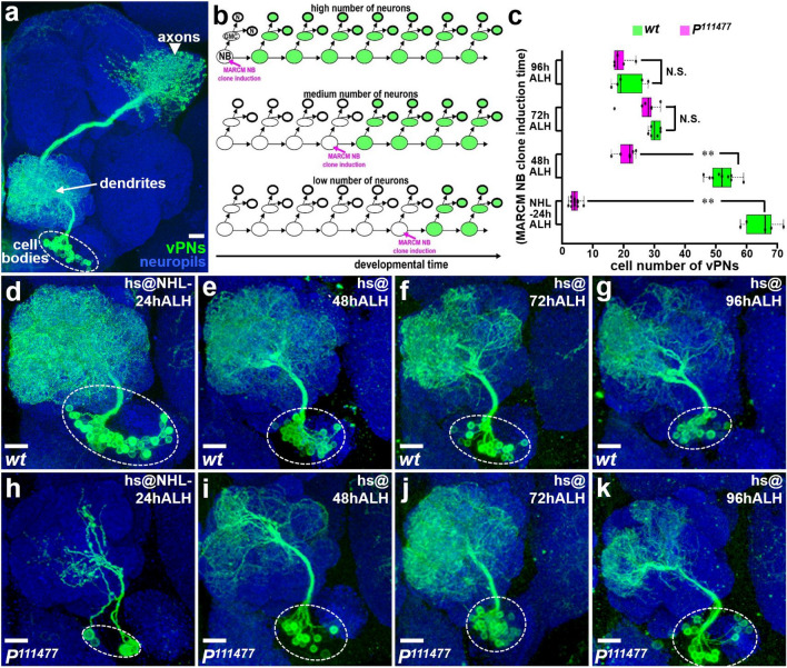
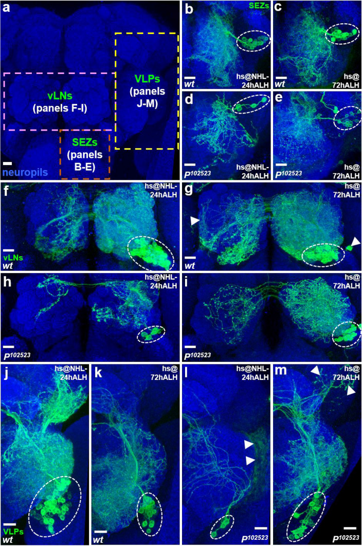
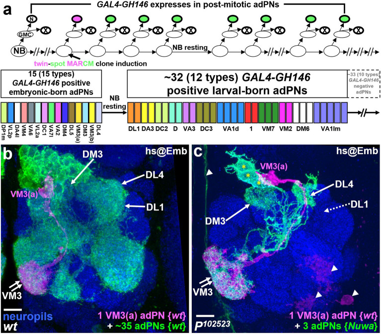
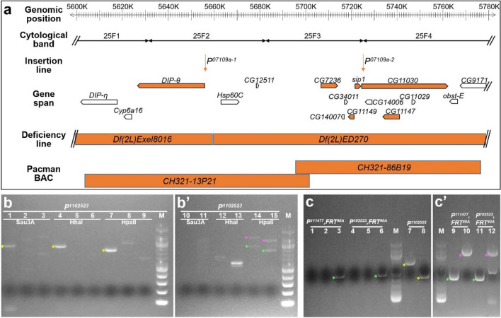
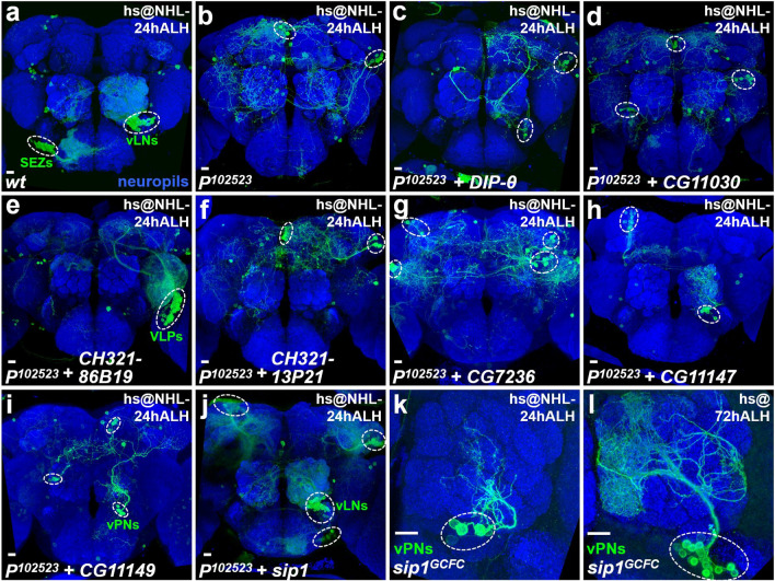
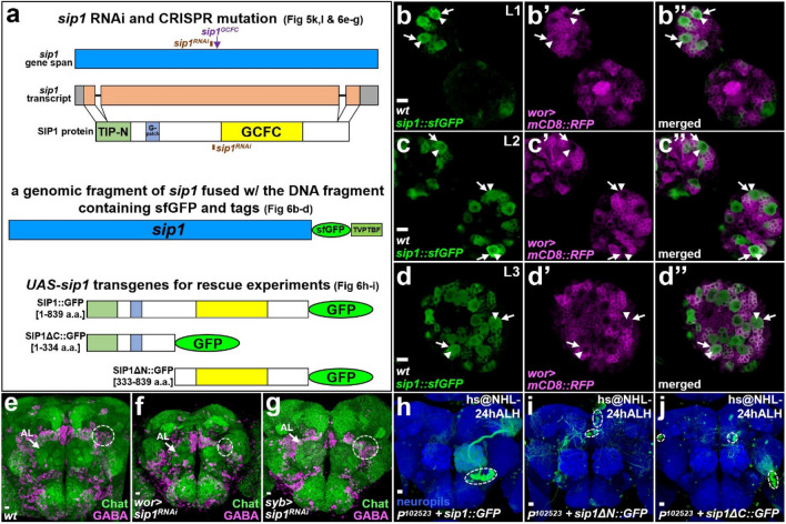
Neurogenesis in the Drosophila central brain progresses dynamically in order to generate appropriate numbers of neurons during different stages of development. Thus, a central challenge in neurobiology is to reveal the molecular and genetic mechanisms of neurogenesis timing. Here, we found that neurogenesis is significantly impaired when a novel mutation, Nuwa, is induced at early but not late larval stages. Intriguingly, when the Nuwa mutation is induced in neuroblasts of olfactory projection neurons (PNs) at the embryonic stage, embryonic-born PNs are generated, but larval-born PNs of the same origin fail to be produced. Through molecular characterization and transgenic rescue experiments, we determined that Nuwa is a loss-of-function mutation in Drosophila septin interacting protein 1 (sip1). Furthermore, we found that SIP1 expression is enriched in neuroblasts, and RNAi knockdown of sip1 using a neuroblast driver results in formation of small and aberrant brains. Finally, full-length SIP1 protein and truncated SIP1 proteins lacking either the N- or C-terminus display different subcellular localization patterns, and only full-length SIP1 can rescue the Nuwa-associated neurogenesis defect. Taken together, these results suggest that SIP1 acts as a crucial factor for specific neurogenesis programs in the early developing larval brain.


Conflict of interest statement

The authors declare no competing interests.

Figures

Figure 1
Figure 1
Neurogenesis of vPNs is significantly impaired when the P111477 mutation is induced at early but not late larval stages. (a) An example of a MARCM neuroblast (NB) clone is used to reveal the morphology of vPNs in the adult brain; using GAL4-MZ699. Axons (arrowhead) were primarily projected to the lateral horn, while dendrites (arrow) were found within the AL, and cell bodies (dashed circle) were distributed ventral to the AL. (b) Schematic drawings show predicted outcomes of MARCM clones induced at different developmental stages: high, medium and low numbers of neurons should be respectively observed when MARCM clones are induced at early, middle and late developmental stages. (c) Numbers of labeled vPNs were shown for wild-type (dg) and P111477 mutants (hk) when MARCM clones were induced at different larval stages. (dk) Gradually reduced numbers of labeled vPNs (within the dashed circle) were observed in wild-type samples (dg) when MARCM clones were induced at 24 h after larval hatching (NHL-24 h ALH), 48 h ALH, 72 h ALH and 96 h ALH. vPN neurogenesis was significantly impaired in the P111477 mutant samples (hk) induced at early but not late larval stages. The genotypes shown in all figures are summarized in Supplemental table 1. Neuropils were revealed by the Bruchpilot (Brp) staining (blue). Scale bar: 10 μm.
Figure 2
Figure 2
Neurogenesis is generally compromised in various brain regions when the Nuwa mutation is induced at the early but not late larval stages. (a) Three groups of neurons were used to examine the developmental stage-dependent requirement for Nuwa, including neurons in the subesophageal zone (SEZs; panels be), ventral olfactory interneurons in the AL (vLNs; panels fi) and neurons in the ventrolateral protocerebrum (VLPs; panels J-M). (bm) All three groups of neurons displayed severe neurogenesis defects with the P102523 (Nuwa) mutation when MARCM clones were induced at NHL-24 h ALH (panels b, d, f, h, j, l), whereas no obvious neurogenesis defects were observed in neurons with the Nuwa mutation when MARCM clones were induced at 72 h ALH (panels c, e, g, i, k, m). Neuropils were revealed by the Brp staining (blue), and background neurons are indicated by arrowheads. Scale bar: 10 μm.
Figure 3
Figure 3
Embryonic-born adPNs are produced normally but postembryonic-born adPNs fail to appear when the Nuwa mutation is induced at the embryonic stage. (a) Schematic drawings show a predicted outcome for a twin-spot MARCM clone when it is induced at the embryonic stage (upper panel); the known cell numbers and subtypes of embryonic- and larval-born adPNs labeled by GAL4-GH146 are shown (bottom panel). (b, c) Examples of wild-type and Nuwa mutant twin-spot MARCM clones induced at the embryonic stage. In the wild-type sample (panel b), a VM3a adPN (an embryonic-born adPN in magenta) was associated with around 35 adPNs (containing both embryonic- and larval-born adPNs in green); in the P102523 (Nuwa) mutant (panel c), a VM3a wild-type adPN (magenta) was associated with three Nuwa mutant adPNs (green, marked by asterisks). Dendrites of the three green Nuwa mutant adPNs were only observed in DM3, VM3 and DL4 [but not DL1 (indicated by dashed arrow)] glomeruli of the AL, indicating that they belong to the last three types of embryonic-born adPNs and further suggesting that no larval-born adPNs were generated. Neuropils were revealed by Brp staining (blue), and background neurons are indicated by arrowheads. Scale bar: 10 μm.
Figure 4
Figure 4
Molecular characterization of Nuwa. (a) Illustration shows genomic position, cytological bands, P-element insertion lines, gene span, deficiency lines and Pacman genomic BAC clones related to Nuwa. Reagents tested in this study are shown in orange. (b, bʹ) Genomic DNA isolated from the P102523 insertion line was cut with restriction enzymes (e.g., Sau3A, HhaI and HpaII), self-ligated into circularized DNAs and then used as a template for inverse PCR. The PCR products contained flanking DNA fragments of P102523, which were mapped to three genes, including Fas3 (yellow asterisks; lanes 1, 4 and 7), DIP-θ (green asterisks; lanes 14 and 15) and CG11030 (magenta asterisks; lanes 14 and 15). (c, cʹ) Genomic DNA isolated from P102523, P111477, FRT40A and P102523, FRT40A mutant lines was directly used for PCR reactions to obtain DNA fragments flanking P-element insertions. P-element-flanking DNA fragments mapped to Fas3 (yellow asterisks) in genomic DNA from the P102523 mutant line (lanes 7 and 8), but not in P111477, FRT40A (lanes 1 and 2) or P102523, FRT40A (lanes 4 and 5) mutant lines. In contrast, P-element-flanking DNA fragments mapped to DIP-θ (green asterisks; lanes 3, 6, 9 and 11) and CG11030 (magenta asterisks; lanes 10 and 12) were found in genomic DNA isolated from P111477, FRT40A and P102523, FRT40A mutant lines. Oligos used for reverse PCR and PCR reactions in panels b, bʹ, c, cʹ are listed in Supplemental table 3.
Figure 5
Figure 5
The Nuwa mutation maps as a loss-of-function of sip1. (ad) Overexpression of the DIP-θ or CG11030 cDNA transgene driven by Act-FRT < stop < FRT-GAL4 failed to rescue the neurogenesis defect in P102523 mutants. All wild-type neural lineages (with cell numbers and morphology) labeled by Act-FRT < stop < FRT-GAL4 can be found in the previous study (Yu et al., 2013; reference #3). (ef) The Nuwa-associated neurogenesis defect in the P102523 mutation was rescued by the CH321-86B19 BAC genomic clone, but not the CH321-13P21 BAC genomic clone. (gi) Overexpression of CG7236, CG111477 and CG11149 cDNA transgenes driven by Act-FRT < stop < FRT-GAL4 still failed to rescue the Nuwa-associated neurogenesis defect in the P102523 mutants. (j) In contrast, overexpression of the sip1 transgene driven by Act-FRT < stop < FRT-GAL4 significantly rescued the neurogenesis defect in the P102523 mutants. (K and L) The custom-made sip1GCFC mutation recapitulated the Nuwa-associated neurogenesis defect. Neuropils were revealed by Brp staining (blue). Scale bar: 10 μm.
Figure 6
Figure 6
The SIP1 expression pattern, RNAi knockdown of sip1 and SIP1:: GFP fusion proteins. (a) Schematic drawing shows the gene span, transcript, protein structure and reagents related to sip1 (upper panel). TIP-N: Tuftelin-interacting protein N terminal domain; G-patch: domain enriched with highly conserved glycines; GCFC: domain containing a sequence similar to a GC-rich sequence DNA-binding factor, transcriptional repressor and histone-interacting proteins. Three SIP1:: GFP fusion proteins were used in this study, including the full-length SIP1:: GFP, SIP1ΔC:: GFP and SIP1ΔN:: GFP (bottom panel). (bd) SIP1:: sfGFP expression (green; the transgenic fly was obtained from Vienna Drosophila Resource Center, VDRC318488) was enriched in neuroblasts. Estimation of the relative SIP1:: sfGFP expression level in neuroblast and neurons can be found in Supplemental Fig. 7. The plasma membrane (outer) and the nuclear membrane (inner) of neuroblasts are indicated by arrows and arrowheads, respectively (labeled in magenta by mCD8:: RFP driven by worniu-GAL4 (wor)). (eg) RNAi knockdown of sip1 using a GAL4 line expressed in neuroblasts, worniu-GAL4 (wor), but not using a pan-neuronal GAL4 line, synaptobrevin-GAL4 (syb), resulted in aberrant brain morphology, including overall smaller brain size, abnormal neuropil architectures (the AL was indicated by arrows). Excitatory and inhibitory neurons were visualized by choline acetyltransferase (Chat; green) and γ-aminobutyric acid (GABA; magenta) staining. A reduction of GABA-positive neuronal number dorsolateral to the AL was observed in wor > sip1RNAi knockdown samples (dashed circles; wild-type: 123.5 ± 13.3, n = 6; wor > sip1RNAi: 39.3 ± 11.3, n = 6; syb > sip1RNAi: 127.3 ± 24.3, n = 6). (hj) The Nuwa-associated neurogenesis defect in the P102523 mutation was rescued by the full-length sip1::GFP transgene, but not sip1ΔN::GFP or sip1ΔC::GFP transgenes. Neuropils were revealed by Brp staining (blue). Scale bar: 10 μm.

Similar articles

References

    1. Urbach R, Technau GM. Molecular markers for identified neuroblasts in the developing brain of Drosophila. Development. 2003;130:3621–3637. doi: 10.1242/dev.00533. - DOI - PubMed
    1. Ito M, Masuda N, Shinomiya K, Endo K, Ito K. Systematic analysis of neural projections reveals clonal composition of the Drosophila brain. Curr. Biol. 2013;23:644–655. doi: 10.1016/j.cub.2013.03.015. - DOI - PubMed
    1. Yu HH, et al. Clonal development and organization of the adult Drosophila central brain. Curr. Biol. 2013;23:633–643. doi: 10.1016/j.cub.2013.02.057. - DOI - PMC - PubMed
    1. Lin S, et al. Extremes of lineage plasticity in the Drosophila brain. Curr. Biol. 2013;23:1908–1913. doi: 10.1016/j.cub.2013.07.074. - DOI - PMC - PubMed
    1. Maurange C, Gould AP. Brainy but not too brainy: starting and stopping neuroblast divisions in Drosophila. Trends Neurosci. 2005;28:30–36. doi: 10.1016/j.tins.2004.10.009. - DOI - PubMed

Publication types

Substances