Meta-Analysis
. 2022 Mar;123(3):601-619.
doi: 10.1002/jcb.30207. Epub 2022 Jan 8.

Iron dysregulation in COVID-19 and reciprocal evolution of SARS-CoV-2: Natura nihil frustra facit

Affiliations
Meta-Analysis

Iron dysregulation in COVID-19 and reciprocal evolution of SARS-CoV-2: Natura nihil frustra facit

Yash Gupta et al. J Cell Biochem. 2022 Mar.

Abstract

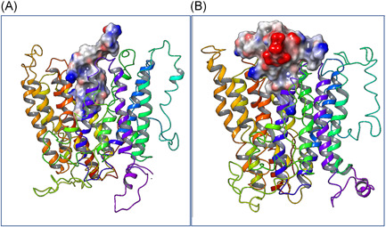
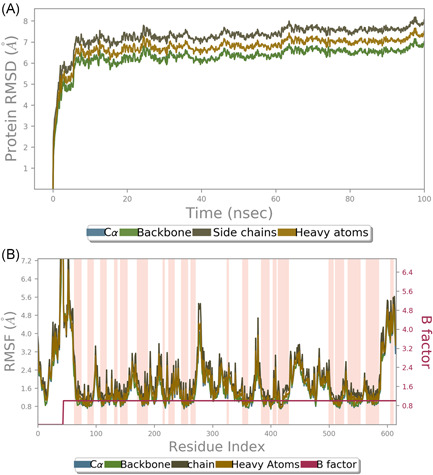
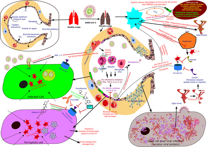
After more than a year of the COVID-19 pandemic, SARS-CoV-2 infection rates with newer variants continue to devastate much of the world. Global healthcare systems are overwhelmed with high positive patient numbers. Silent hypoxia accompanied by rapid deterioration and some cases with septic shock is responsible for COVID-19 mortality in many hospitalized patients. There is an urgent need to further understand the relationships and interplay with human host components during pathogenesis and immune evasion strategies. Currently, acquired immunity through vaccination or prior infection usually provides sufficient protection against the emerging variants of SARS-CoV-2 except Omicron variant requiring recent booster. New strains have shown higher viral loads and greater transmissibility with more severe disease presentations. Notably, COVID-19 has a peculiar prognosis in severe patients with iron dysregulation and hypoxia which is still poorly understood. Studies have shown abnormally low serum iron levels in severe infection but a high iron overload in lung fibrotic tissue. Data from our in-silico structural analysis of the spike protein sequence along with host proteolysis processing suggests that the viral spike protein fragment mimics Hepcidin and is resistant to the major human proteases. This functional spike-derived peptide dubbed "Covidin" thus may be intricately involved with host ferroportin binding and internalization leading to dysregulated host iron metabolism. Here, we propose the possible role of this potentially allogenic mimetic hormone corresponding to severe COVID-19 immunopathology and illustrate that this molecular mimicry is responsible for a major pathway associated with severe disease status. Furthermore, through 3D molecular modeling and docking followed by MD simulation validation, we have unraveled the likely role of Covidin in iron dysregulation in COVID-19 patients. Our meta-analysis suggests the Hepcidin mimetic mechanism is highly conserved among its host range as well as among all new variants to date including Omicron. Extensive analysis of current mutations revealed that new variants are becoming alarmingly more resistant to selective human proteases associated with host defense.

Keywords: MD simulations; evolved variants; ferritin-transferrin paradox; host proteases; hypoxia; iron homeostasis.


Conflict of interest statement

The authors declare that there are no conflict of interests.

Figures

Figure 1
Figure 1
Phylogenetic analysis of Hepcidin hormone reveals a grouping among mammals with severe SARS‐CoV‐2 infection, while the peptide in primates and bats seems to be more evolved. Still, the similarity between the two groups is surprising due to the actual species evolutionary distance between the two
Figure 2
Figure 2
Multiple sequence alignment of spike fragment homologous to Hepcidin we now call Covidin with different mammals. The hotter regions (toward the red spectrum) are highly conserved while colder (toward the blue spectrum) are the least conserved. Hepcidin seems to be highly conserved among all mammals and has high homology with the Covidin. Interestingly, the Covidin homology is higher for pangolin and bat which are postulated to be viral reservoirs suggesting an evolutionary advantage conferred by this peptide for viral pathogenesis
Figure 3
Figure 3
Current mutation rates of different regions of the SARS‐CoV‐2 genome (GSAID‐Nextstarain). Data from 4298 genomes sampled between December 2019 and August 2021 shows the Covidin peptide has 0.0028 average mutational diversity making it highly conserved compared to 0.2 and 0.05 average mutational diversity for whole‐genome and spike protein, respectively. Mutations are represented by vertical bars with sizes proportional to percent frequencies. (A) Genome‐wide mutation rates, (B) mutation rates in Spike protein, and (C) mutation rates in the Covidin region
Figure 4
Figure 4
The protease map of Spike protein (N terminal on the left and C terminal on the right). This analysis suggests that if a spike protein is degraded by human protease, there is a high probability of releasing Hepcidin like a peptide fragment, that is, Covidin. This spike degradation could result following normal endosomal degradation of the spike, post nucleocapsid delivery in the cytoplasm, or necrosis of the dead virus‐infected cell with the unassembled surplus spike. The sequence of the peptide at position 1214‐1255 with respect to Ref:UniProt “P0DTC2” (Wuhan isolate) is “N‐term‐‘WYIWLGFIAGLIAIVMVTIMLCCMTSCCSCLKGCCSCGSCCK’‐C‐term”
Figure 5
Figure 5
Procleave prediction of variation in protease site scores due to mutations in various variant spike sequences (Table 1) relative to the Wuhan sequence mapped on 3D structure (PDB ID: 7KJ2). Proteases act as innate immune components by degrading foreign peptides thus rendering them ineffective. The variant mutations, though mainly on the protein surface, do not seem to confer much immunological advantage, as only a few seem to improve receptor binding. Mutations have long been attributed to greater stability and transmissibility of viruses, in part by enabling the survival of proteases present in the host system
Figure 6
Figure 6
Schematic representation of Comparative 3D structure of interacting (A) Natural Hepcidin and (B) Covidin docked at the central pore of ferroportin
Figure 7
Figure 7
(A) Comparative amino acid interaction map of Hepcidin versus Covidin with Ferroportin central pore extracellular domains. The colored interactions are host Hepcidin with Ferroportin and grayscale are Covidin with ferroportin in the same space. (B) Comparative amino acid interaction map of Covidin versus Hepcidin with Ferroportin central pore extracellular domains. The colored interactions are Covidin with Ferroportin and grayscale are Hepcidin with ferroportin in the same space. Both Hepcidin and Covidin bind strongly to the central pore from the extracellular space of a host iron transporter “ferroportin” and the resulting complex has similar interaction features and binding space as the natural hormone Hepcidin
Figure 8
Figure 8
MD simulation (100 ns) of natural Hepcidin hormone bound to ferroportin. The docking was highly stable with negligible deviation from original throughout the simulation and the Orientations of Proteins in Membranes predicted membrane topology chosen for simulation was also highly stable
Figure 9
Figure 9
MD simulation (100 ns) of Covidin viral origin peptide bound to ferroportin. The docking was highly stable with negligible deviation from the original dock throughout the simulation, and the Orientations of Proteins in Membranes predicted membrane topology chosen for simulation was also highly stable
Figure 10
Figure 10
Cartoon representation of overlap of hypothetical iron dysregulation by Hepcidin‐like peptide Covidin and the actual clinical picture of COVID‐19 patients. The lung fibrosis observed in COVID‐19 patients and resulting hypoxia is the main reason for mortality in severe cases now seconded by secondary bacterial and fungal infections. Classic pathways do not govern COVID‐19 associated lung fibrosis and a deviation has been reported by multiple workers. Ferritin is shown to be highly expressed in COVID‐19 patients due to inflammation mediators such as IL‐6 and cytokine storm or increased release from damaged/ferroptosis cells, contrastingly the serum iron and transferrin levels are very low. Sphingosine‐1 is one of the markers of severe COVID‐19 is induced by high serum iron or ferroptosis. But as severe COVID‐19 is accompanied by hypoxia which strongly reduces Hepcidin levels through erythropoietin (EPO) hormone, such iron dysregulation can be attributed to Covidin peptide which we have found to be proteolysis resistant against common and major human proteases and thus can be present in very high concentrations once excess spike protein is phagocytosed in dying infected cells. Hypoxia is essential in escalating viral infection, and it induces surface localization of Furin protease bypassing the endosomal route of nucleocapsid but rather plasma membrane fusion by cell surface spike processing by furin enzyme. Also, SARS‐CoV‐2 and hypoxia‐induced ACE2 overexpression should repress TGF‐β and reduce fibrosis, the opposite of what is observed. Our proteolysis, protein modeling, peptide docking, and MD simulation experiments strongly support functional biological mimicry of Covidin with the natural host Hepcidin hormone. Further, this association seems to be more conserved with the proposed primary hosts of SARS‐CoV‐2, supporting evidence of high iron requirement by relatively large and resource‐intensive high viral turnover of SARS‐CoV‐2. Further, Vitamin D and Mn2+ have been effective in reducing the severity, and in the case of Vit. D, reduced mortality as seen in large trials. However, the mechanism for which is still not well established. As these agents induce overexpression of ferroportin, the reduction of ferroptosis and thereby Sphingosine‐1 mediated fibrosis could be a plausible mechanism of action for observed protection. This can be further coupled with Hepcidin hormone antagonists like Fursultiamine (FDA approved vitamin supplement) or LY3127804 (monoclonal antibody, Phase2) to reverse the severe fibrosis and possibly ferroptosis in lung alveoli epithelia. As Hepcidin/Covidin causes ferroportin degradation by ubiquitin proteasomal pathway, antagonists alone cannot reverse the intracellular iron overload. Reduced erythropoiesis due to low serum iron might also accelerate hypoxia, rapidly deteriorating patient condition, and aggravating COVID‐19
Figure 11
Figure 11
Overview of the deciphered relationship of different subsets of COVID‐19 pathology and evolution

Comment in

References

    1. Gupta Y, Maciorowski D, Zak SE, et al. Bisindolylmaleimide IX: a novel anti‐SARS‐CoV2 agent targeting viral main protease 3CLpro demonstrated by virtual screening pipeline and in‐vitro validation assays. Methods. 2021a;195:57‐71. 10.1016/j.ymeth.2021.01.003 - DOI - PMC - PubMed
    1. Lauer SA, Grantz KH, Bi Q, et al. The incubation period of coronavirus disease 2019 (COVID‐19) from publicly reported confirmed cases: estimation and application. Ann Intern Med. 2020;172(9):577‐582. - PMC - PubMed
    1. COVID C, Team R. SARS‐CoV‐2 B. 1.1. 529 (Omicron) variant—United States, December 1–8, 2021. MMWR Morb Mortal Wkly Rep. 2021;70(50):1731‐1734. - PMC - PubMed
    1. Homma Y, Katsuta T, Oka H, et al. The incubation period of the SARS‐CoV‐2 B. 1.1. 7 variant is shorter than that of other strains. J Infect. 2021;83:e15‐e17. 10.1016/j.jinf.2021.06.011 - DOI - PMC - PubMed
    1. Snell LB, Awan AR, Charalampous T, et al. SARS‐CoV‐2 variants with shortened incubation periods necessitate new definitions for nosocomial acquisition. J Infect. 2021. 10.1016/j.jinf.2021.08.041 - DOI - PMC - PubMed

Publication types

Supplementary concepts