Review
. 2022 Feb;39(2):339-370.
doi: 10.1111/echo.15266. Epub 2022 Jan 7.

Echocardiography for extracorporeal membrane oxygenation

Affiliations
Review

Echocardiography for extracorporeal membrane oxygenation

Patrick T Hussey et al. Echocardiography. 2022 Feb.

Abstract

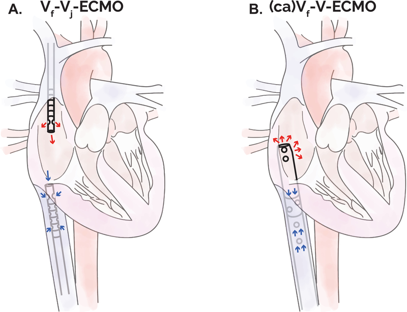
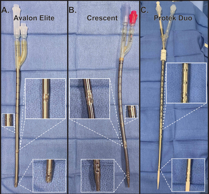
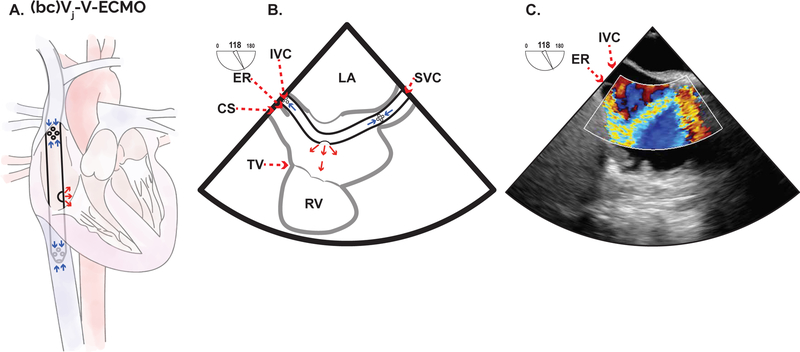
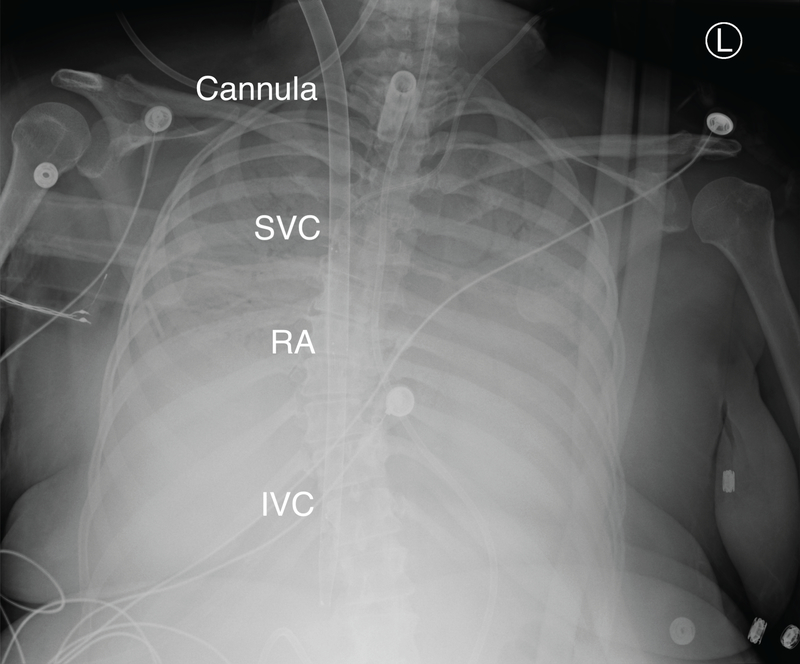
Extracorporeal membrane oxygenation (ECMO) provides advanced cardiopulmonary life support for patients in cardiac and/or respiratory failure. Echocardiography provides essential diagnostic and anatomic information prior to ECMO initiation, allows for safe and efficient ECMO cannula positioning, guides optimization of flow, provides a modality for rapid troubleshooting and patient evaluation, and facilitates decision-making for eventual weaning of ECMO support. Currently, guidelines for echocardiographic assessment in this clinical context are lacking. In this review, we provide an overview of echocardiographic considerations for advanced imagers involved in the care of these complex patients. We focus predominately on new cannulas and complex cannulation techniques, including a special focus on double lumen cannulas and a section discussing indirect left ventricular venting. Echocardiography is tremendously valuable in providing optimal care in these challenging clinical situations. It is imperative for imaging physicians to understand the pertinent anatomic considerations, the often complicated physiological and hemodynamic context, and the limitations of the imaging modality.

Keywords: LAVA-ECMO; VA-ECMO; VV-ECMO; dual-lumen cannulation; echocardiography; extracorporeal membrane oxygenation.


Figures

Figure 1.
Figure 1.. Schematic of common VV-ECMO cannulation strategies.
The Maastricht consensus nomenclature is used to describe cannula location. “Traditional” two-cannula approach to VV-ECMO. In Vf-Vj-ECMO the femoral venous drainage cannula tip is positioned within the IVC and an internal jugular venous return cannula tip is positioned within the right atrium (A). In (ca)Vf-V-ECMO a single, double-lumen (cavoatrial) cannula is placed with venous return achieved from the IVC and return flow into the RA. One example is the ProtekDuo RD cannula (CardiacAssist) (B). A similar strategy may be achieved with two separate femoral venous cannulas. ca: cavoatrial, ECMO: extracorporeal membrane oxygenation, f: femoral, IVC: inferior vena cava, j: jugular, RA: right atrium, SVC: superior vena cava, VV: veno-venous.
Figure 2.
Figure 2.. Examples of double lumen ECMO cannulas.
The Avalon Elite (Getinge AB) (A) and Crescent (MC3) (B) are used for bicaval cannulation with two inflow ports designed to be positioned within the SVC and IVC and a central outflow port that is oriented to direct flow towards the center of the tricuspid valve. The Protek Duo (CardiacAssist) has proximal inflow ports with outflow ports at the distal tip of the cannula (C). These cannulas come in various lengths and can be placed from the internal jugular vein and positioned with the proximal inflow port within the RA and the distal outflow port within the PA. Alternatively, the femoral vein can be cannulated, the proximal inflow ports positioned within the IVC, and the distal outflow port oriented within the RA. A shorter option, the Protek Duo RD cannula is pictured. ECMO: extracorporeal membrane oxygenation, IVC: inferior vena cava, RA: right atrium, SVC: superior vena cava, PA: pulmonary artery.
Figure 3.
Figure 3.. Double lumen bicaval cannulation for VV-ECMO.
A common method for conducting VV-ECMO is through placement of a double lumen cannula via the right internal jugular vein [(bc)Vj-V-ECMO]. Inflow ports are then positioned within the IVC and the SVC and a central outflow port is then oriented within the RA to direct blood flow across the tricuspid valve (A). Examples include the Avalon Elite (Getinge AB) and the Crescent (MC3). TEE readily confirms appropriate outflow port positioning and outflow jet orientation towards the tricuspid valve using color Doppler imaging. The mid-esophageal modified bicaval view is useful during positioning (B) and is often found by increasing the omniplane angle and making a slight clockwise turn of the probe from the mid-esophageal bicaval view. A modified bicaval view of a Crescent dual-lumen cannula with color flow Doppler demonstrating an outflow jet appropriately directed towards the center of the TV (C). bc: bicaval, CS: coronary sinus, ER: Eustachian ridge, IVC: inferior vena cava, LA: left atrium, RA: right atrium, SVC: superior vena cava, TV: tricuspid valve, VV-ECMO: veno-venous extracorporeal membrane oxygenation.
Figure 4.
Figure 4.. Radiographic appearance of dual lumen veno-venous ECMO cannula.
A Crescent MC3 (Cardiopulmonary, Dexter, MI) bicaval dual lumen cannula in a patient on (bc)Vj-V-ECMO support. The radiopaque markers at the site of SVC drainage, the infusion site (RA), the site of IVC drainage, and the catheter tip are readily apparent. (bc): bicaval, ECMO: extracorporeal membrane oxygenation, IVC: inferior vena cava, Vj: venous (jugular), RA: right atrium, SVC: superior vena cava.
Figure 5.
Figure 5.. Protek Duo cannulation for temporary RV support.
Double lumen cannulas are available for mechanical support of the RV including the Protek Duo (CardiacAssist) and the Impella RP (Abiomed). The Protek Duo is placed via the IJ with the inflow ports positioned within the RA and the distal outflow in the main PA [(dl)Vj-P-ECMO] allowing for offloading of the RV (A). The midesophageal RV inflow-outflow can demonstrate the Protek Duo cannula traversing the TV and the tip and outflow port location distal to the PV (B). A TEE view with color flow Doppler demonstrating outflow from the distal port within the main PA (C). dl: double lumen, ECMO: extracorporeal membrane oxygenation, j:jugular, P: Pulmonary artery, PV: Pulmonic valve, RV: right ventricle.
Figure 6.
Figure 6.. Percutaneous, transseptal indirect left ventricular venting.
To address LV distention during VA-ECMO and indirect LV venting strategy may be employed. One approach is placement of a dedicated transseptal cannula across the IAS to provide LA drainage and consequent indirect LV venting. Alternatively, a single, multistage cannula may be employed for percutaneous transseptal cannulation with the distal inflow ports positioned within the LA and the proximal inflow ports within the inferior cavoatrial junction (A). As depicted (A) this represents both femoral venous drainage and left atrial venting in addition to the arterial return via the femoral artery. Purple arrows represent return of blood with variable oxygenation contingent upon the clinical scenario. A non-standard mid or upper esophageal window can demonstrate the position of a transseptal cannula and the inflow ports and can identify key intracardiac structures including the IAS, the RPA, and the AscAo (B). A TEE view demonstrating color Doppler inflow of blood into a multistage venous cannula positioned across the IAS (C). al: left atrium, AscAo: Ascending aorta, ECMO: Extracorporeal membrane oxygenation, f: Femoral, IAS: Interatrial septum, IVC: Inferior vena cava, LA: Left atrium, LV: Left ventricle, RA: Right atrium, RPA: Right pulmonary artery.

Similar articles

Cited by

References

    1. Grant C Jr., Richards JB, Frakes M, Cohen J, Wilcox SR. ECMO and Right Ventricular Failure: Review of the Literature. J Intensive Care Med. 2021;36(3):352–360. - PubMed
    1. Bouferrache K, Vieillard-Baron A. Acute respiratory distress syndrome, mechanical ventilation, and right ventricular function. Curr Opin Crit Care. 2011;17(1):30–35. - PubMed
    1. Hahn RT, Abraham T, Adams MS, et al. Guidelines for performing a comprehensive transesophageal echocardiographic examination: recommendations from the American Society of Echocardiography and the Society of Cardiovascular Anesthesiologists. J Am Soc Echocardiogr. 2013;26(9):921–964. - PubMed
    1. Doufle G, Roscoe A, Billia F, Fan E. Echocardiography for adult patients supported with extracorporeal membrane oxygenation. Crit Care. 2015;19:326. - PMC - PubMed
    1. Rudski LG, Lai WW, Afilalo J, et al. Guidelines for the echocardiographic assessment of the right heart in adults: a report from the American Society of Echocardiography endorsed by the European Association of Echocardiography, a registered branch of the European Society of Cardiology, and the Canadian Society of Echocardiography. J Am Soc Echocardiogr. 2010;23(7):685–713; quiz 786–688. - PubMed