. 2022 May;94(5):2019-2025.
doi: 10.1002/jmv.27577. Epub 2022 Jan 19.

The puzzling mutational landscape of the SARS-2-variant Omicron

Affiliations

The puzzling mutational landscape of the SARS-2-variant Omicron

Jacques Fantini et al. J Med Virol. 2022 May.

Abstract

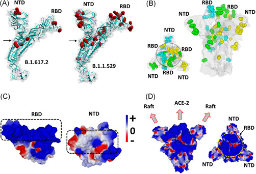
The recently emerging SARS-CoV-2 variant omicron displays an unusual association of 30 mutations, 3 deletions, and 1 insertion. To analyze the impact of this atypic mutational landscape, we constructed a complete structure of the omicron spike protein. Compared with the delta variant, the receptor-binding domain (RBD) of omicron has an increased electrostatic surface potential, but a decreased affinity for the ACE-2 receptor. The N-terminal domain (NTD) has both a decreased surface potential and a lower affinity for lipid rafts. The omicron variant is predicted to be less fusogenic and thus less pathogenic than delta, due to a geometric reorganization of the S1-S2 cleavage site. Overall, these virological parameters suggest that omicron does not have a significant infectivity advantage over the delta variant. However, in omicron, neutralizing epitopes are greatly affected, suggesting that current vaccines will probably confer little protection against this variant. In conclusion, the puzzling mutational pattern of the omicron variant combines contradictory properties which may either decrease (virological properties) or increase (immunological escape/facilitation) the transmission of this variant in the human population. This Janus-like phenotype may explain some conflicting reports on the initial assessment of omicron and provide new insights about the molecular mechanisms controlling its dissemination and pathogenesis worldwide.

Keywords: SARS coronavirus; antibody susceptibility; coronavirus; evolution; infection; pathogenesis; virulence; virus classification.


Conflict of interest statement

Didier Raoult has a conflict of interest being a consultant for Hitachi High‐Technologies Corporation, Tokyo, Japan from 2018 to 2020. All other authors have no conflicts of interest to declare. Funding sources had no role in the design and conduct of the study; collection, management, analysis, and interpretation of the data; and preparation, review, or approval of the manuscript.

Figures

Figure 1
Figure 1
Structural analysis of omicron spike protein. (A) Molecular models of the delta B.1.617.2 and omicron B.1.1.529 spike proteins with mutations highlighted in red and the S1‐S2 cleavage site indicated by an arrow. (B) Spike trimer of the omicron variant as viewed from the host cell surface (left panel, central RBDs indicated within a circle), or perpendicularly to the virus envelope (right panel). Mutations are colored in cyan, green and yellow corresponding, respectively, to spike subunits A, B, and C. The NTD and RBD of each chain is indicated. The protein surface is colored in grey. (C) Electrostatic surface potential of omicron RBD and NTD. The color scale for the electrostatic surface potential (negative in red, positive in blue, neutral in white) is indicated. The regions of the RBD and the NTD, respectively, bound to ACE‐2 and the lipid raft are indicated by dashed rectangles. (D) Two views of the electrostatic surface potential of the trimeric spike of omicron (left panel, lateral view of the trimer perpendicular to viral envelope; right panel, top view of the trimer facing the host cell surface, the central RBDs are indicated by a yellow circle). NTD, N‐terminal domain; RBD, receptor‐binding domain
Figure 2
Figure 2
T‐index and I‐index analysis of SARS‐CoV‐2 variants. T‐index values of Wu (Wuhan strain), alpha (α), beta (β), and gamma (γ) were from ref. I‐index values of Wu (Wuhan strain), alpha (α), beta (β), and gamma (γ) were from ref. The T‐index and I‐index of delta (δ) and omicron (ο) were calculated in the present study
Figure 3
Figure 3
Scanning electron microscopy of a SARS‐CoV‐2 Omicron‐RNA‐positive culture supernatant. Image was obtained using a SU5000 scanning electron microscope (SEM) (Hitachi High‐Technologies Corporation). A viral particle is indicated by a white arrow

Comment in

  • Why SARS-CoV-2 vaccination still matters in Africa.
    Taylor-Robinson SD, Morgan MY, Spearman CW, Suliman AAA, Corrah T, Oleribe OO, Taylor-Robinson AW. Taylor-Robinson SD, et al. QJM. 2022 Mar 22;115(3):191-192. doi: 10.1093/qjmed/hcac014. QJM. 2022. PMID: 35080615 No abstract available.

References

    1. Callaway E. Heavily mutated Omicron variant puts scientists on alert. Nature. 2021;600:21. - PubMed
    1. He X, Hong W, Pan X, Lu G, Wei X. SARS‐CoV‐2 Omicron variant: characteristics and prevention. MedComm. 2021;2:838‐845. - PMC - PubMed
    1. Fantini J, Yahi N, Azzaz F, Chahinian H. Structural dynamics of SARS‐CoV‐2 variants: a health monitoring strategy for anticipating Covid‐19 outbreaks. J Infect. 2021;83:197‐206. - PMC - PubMed
    1. Jaafar R, Boschi C, Aherfi S. High individual heterogeneity of neutralizing activities against the original strain and nine different variants of SARS‐CoV‐2. Viruses. 2021;13(11):2177. - PMC - PubMed
    1. Tian D, Sun Y, Zhou J, Ye Q. The global epidemic of the SARS‐CoV‐2 delta variant, key spike mutations and immune escape. Front Immunol. 2021;12:751778. - PMC - PubMed

Publication types

Supplementary concepts