. 2022 Jan 8;20(1):4.
doi: 10.1186/s12964-021-00798-9.

Functional signaling test identifies HER2 negative breast cancer patients who may benefit from c-Met and pan-HER combination therapy

Affiliations

Functional signaling test identifies HER2 negative breast cancer patients who may benefit from c-Met and pan-HER combination therapy

Ian A MacNeil et al. Cell Commun Signal. .

Abstract

Background: Research is revealing the complex coordination between cell signaling systems as they adapt to genetic and epigenetic changes. Tools to uncover these highly complex functional linkages will play an important role in advancing more efficacious disease treatments. Current tumor cell signal transduction research is identifying coordination between receptor types, receptor families, and transduction pathways to maintain tumor cell viability despite challenging tumor microenvironment conditions.

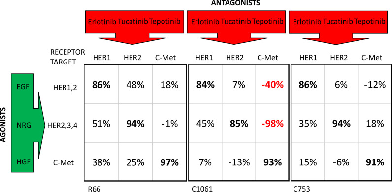
Methods: In this report, coactivated abnormal levels of signaling activity for c-Met and HER family receptors in live tumor cells were measured by a new clinical test to identify a subpopulation of breast cancer patients that could be responsive to combined targeted therapies. The CELsignia Multi-Pathway Signaling Function (CELsignia) Test uses an impedance biosensor to quantify an individual patient's ex vivo live tumor cell signaling response in real-time to specific HER family and c-Met co-stimulation and targeted therapies.

Results: The test identified breast tumors with hyperactive HER1, HER2, HER3/4, and c-Met coordinated signaling that express otherwise normal amounts of these receptors. The supporting data of the pre-clinical verification of this test included analyses of 79 breast cancer patients' cell response to HER and c-Met agonists. The signaling results were confirmed using clinically approved matching targeted drugs, and combinations of targeted drugs in addition to correlative mouse xenograft tumor response to HER and c-Met targeted therapies.

Conclusions: The results of this study demonstrated the potential benefit of a functional test for identifying a subpopulation of breast cancer patients with coordinated abnormal HER and c-Met signaling for a clinical trial testing combination targeted therapy. Video Abstract.

Keywords: Combination targeted therapy; Dysfunctional signaling; HER; c-Met.


Conflict of interest statement

All authors are employed by Celcuity, Inc. 16305 36th Ave N, Suite 100, Minneapolis, MN 55446.

Figures

Fig. 1
Fig. 1
Combination of pan-HER and c-Met inhibitors effectively block the HER-family and the c-Met receptor coactivated functions. Data for an example patient, C135, demonstrates the efficacy of combining neratinib and tepotinib against abnormal stimulation response generated by simultaneous application of the three growth factors (GF)—NRG, EGF, and HGF. Each curve is labeled in the figure to indicate the inhibitor(s) that was added to different wells containing the same number of cells. The order of efficacy response compared to the growth factor only well (at the top of the graph) from low response to high is as follows—Tepotinib alone is least efficacious, Neratinib alone is more efficacious, and the most efficacious treatment is the combination of neratinib and tepotinib data curve that in the figure is just above the dotted blue baseline for untreated cells (no agonist, no antagonist control)
Fig. 2
Fig. 2
Clinical test procedure and c-Met cutoff determination and prevalence analysis using the CELsignia test in a population of HER2 negative breast cancer patients. a Clinical test procedure summary: A 14-gauge to 18-gauge needle biopsy containing about 25 mg of tumor tissue is removed and sent by overnight courier to the Celcuity lab in Minneapolis. After a brief, no passage, culture establishes the viability of the tumor cells, a CELsignia multipathway test is performed using an impedance-based biosensor (96-well format device). Typically, an abnormal signaling report is returned to the requesting physician in less than 14 days. b CELsigniaTest Score distribution density analysis: The likelihood ratio test for the number of components gave the following parameters. For the three-component model, the components 2 (purple) & 3 (blue) means are more than four standard deviations apart. c ROC curve for the three-component fit plots sensitivity versus false positive rate for various cut-off points. Formal significance testing shows that a two-component mixture fits much better than a common normal distribution, and a three-component mixture fits better than two. d False positive rate (blue line) and sensitivity (black line) are presented as a function of the cutoff used
Fig. 3
Fig. 3
HER2 and c-Met receptor are not overexpressed on test tumor cells. a Flow cytometry analysis of HER2/HER3 expression confirmed the HER2- status designated by IHC and/or ISH. Seventy-nine patient samples were analyzed for HER2 and HER3 expression using flow cytometry as described in the methods. The mean fluorescence intensity (MFI) of the samples were compared to three cell lines used as standards in the DAKO HercepTest. (SKBR3-DAKO 3+, MDA175vii- DAKO 1+, MDA231-DAKO 0). Size of the circles indicates the HER2 signaling CELsignia score. b Comparison of expression levels of HGF receptor (c-Met) to HGF response on 79 patient samples using flow cytometry as described in the methods. The size of the circles in both panels indicates the HER2 signaling CELsignia score
Fig. 4
Fig. 4
Determining the IC50 values of pan-HER and c-MET inhibitors from a patient-derived sample (Patient R66) a Percent response of stimulation of NRG1b/EGF signal (two-growth factor cocktail) when treated with a 5-point, 1000-fold titration of the six indicated pan-HER antagonists, exponential log-scale fit with GraphPad Prism. b Percent response of stimulation of HGF signal when treated with a 5-point, 1000-fold dose response of the five indicated c-Met antagonists
Fig. 5
Fig. 5
Cross-talk analysis of individual antagonists against EGF, NRG1b, and c-Met illustrates receptor signaling co-involvement. Percent inhibitions of abnormal signaling are listed for paired antagonists (Tables 5, 6, 7) . The data in Fig. 5 demonstrates for three different HER2 negative patients where treatment with the antagonist drug indicated at the column head affects the signaling generated by the agonist indicated at the matching row for each patient. For ‘on target’ pairing of drug and agonist for each patient shown, the data for duplicate wells demonstrate > 85% efficacy at reducing the abnormal signaling (indicated in bold font) directly related to the target binding receptor indicated in column headings. When considering ‘off target’ effects for patient C1061 for example, Tepotinib treatment leads to a 98% increase in signaling (negative value for inhibition, in red font) upon NRG addition and 40% increase for EGF addition. For C1061 and patient C753, treatment with a HER2 specific antagonist leads to an increase in HGF signaling
Fig. 6
Fig. 6
CELsignia test is a more sensitive and rapid test for detecting drug efficacy compared to the biological correlates. HCC1954 cells were seeded in collagen-fibronectin (CF) coated culture plates and treated 6 h later with tepotinib (250 nM) and neratinib (250 nM), either singly or in combination, as indicated. Cells were harvested and then analyzed by flow cytometry for the markers shown following a period of 16 h (a) or 48 h (b) of drug treatment. The data indicate relative insensitivity of flow cytometry markers to deteriorating cell health until at least 48 h after drug application. At 48 h, annexin v expression has increased by nearly twofold for cells treated with combined pan-HER and cMET targeted drugs
Fig. 7
Fig. 7
Single-cell analysis of AKT and FAK pathway activation does not reveal the HER-dependent differential sensitivity or drug synergy of HCC1954 cells to pan-HER and c-MET inhibitor combinations identified by CELsignia. HCC1954 cells were treated with c-MET (tepotinib) and pan-HER (neratinib) inhibitors, either singly or in combination, at the doses indicated for 16 h as described in Fig. 6 and then analyzed by intracellular phospho-flow cytometry. a, b The mean and standard deviation of levels of pAKT and pFAK in gated live cells following drug treatment from two experiments is shown. c Cells were treated with increasing doses of tepotinib or neratinib, administered singly or in combination as indicated, and analyzed for pAKT and pFAK expression patterns within single cells. At least 10,000 events were acquired per sample and data shown is representative of two experiments
Fig. 8
Fig. 8
Combination of pan-HER and c-Met inhibitors effectively reduce tumor size in the xenograft model. Activity of c-MET and HER specific antagonists in a HCC1954 a NSG mouse xenograft model. Ten mice cohorts were dosed orally QD for 21 days. Mouse weights (Panels A and C) and tumor size (Panels B and D) were recorded every 3 days

Similar articles

Cited by

References

    1. Wagner JP, Wolf-Yadlin A, Sevecka M, Grenier JK, Root DE, Lauffenburger DA, et al. Receptor tyrosine kinases fall into distinct classes based on their inferred signaling networks. Sci Signal. 2013;6(284):ra58. - PMC - PubMed
    1. Viticchie G, Muller PAJ. c-Met and other cell surface molecules: interaction. Activ Funct Conseq Biomed. 2015;3(1):46–70. - PMC - PubMed
    1. Watson SS, Dane M, Chin K, Tatarova Z, Liu M, Liby T, et al. Microenvironment-mediated mechanisms of resistance to HER2 inhibitors differ between HER2+ breast cancer subtypes. Cell Syst. 2018;6(3):329–42. - PMC - PubMed
    1. Liu X, Wang Q, Yang G, Marando C, Koblish HK, Hall LM, et al. A novel kinase inhibitor, INCB28060, blocks c-MET-dependent signaling, neoplastic activities, and cross-talk with EGFR and HER-3. Clin Cancer Res. 2011;17(22):7127–7138. - PubMed
    1. Liu X, Zhao L, Derose YS, Lin YC, Bieniasz M, Eyob H, et al. Short-form ron promotes spontaneous breast cancer metastasis through interaction with phosphoinositide 3-kinase. Genes Cancer. 2011;2(7):753–762. - PMC - PubMed

Publication types

Substances