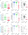
Genotype-specific features reduce the susceptibility of South American yellow fever virus strains to vaccine-induced antibodies
- PMID: 34998466
- PMCID: PMC10067022
- DOI: 10.1016/j.chom.2021.12.009
Genotype-specific features reduce the susceptibility of South American yellow fever virus strains to vaccine-induced antibodies
Abstract
The resurgence of yellow fever in South America has prompted vaccination against the etiologic agent, yellow fever virus (YFV). Current vaccines are based on a live-attenuated YF-17D virus derived from a virulent African isolate. The capacity of these vaccines to induce neutralizing antibodies against the vaccine strain is used as a surrogate for protection. However, the sensitivity of genetically distinct South American strains to vaccine-induced antibodies is unknown. We show that antiviral potency of the polyclonal antibody response in vaccinees is attenuated against an emergent Brazilian strain. This reduction was attributable to amino acid changes at two sites in central domain II of the glycoprotein E, including multiple changes at the domain I-domain II hinge, which are unique to and shared among most South American YFV strains. Our findings call for a reevaluation of current approaches to YFV immunological surveillance in South America and suggest approaches for updating vaccines.
Keywords: 17D; South America; antibody response; emerging virus; flavivirus; glycoprotein; immunological response; neutralizing antibodies; vaccine; yellow fever virus.
Copyright © 2021 Elsevier Inc. All rights reserved.
Conflict of interest statement
Declaration of interests K.C. is a member of the scientific advisory boards of Integrum Scientific, Biovaxys Technology, and the Pandemic Security Initiative of Celdara Medical. A.Z.W., M.S., and L.M.W. are/were employees of Adimab, and may hold shares in Adimab. L.M.W. is an employee of Adagio Therapeutics, and holds shares in Adagio Therapeutics. C.L.M. and Z.A.B. are employees of Mapp Biopharmaceutical.
Figures


Comment in
-
Even old foes can learn sweet new tricks.Cell Host Microbe. 2022 Feb 9;30(2):151-153. doi: 10.1016/j.chom.2022.01.011. Cell Host Microbe. 2022. PMID: 35143767
Similar articles
-
Evaluation of humoral immune response after yellow fever infection: an observational study on patients from the 2017-2018 sylvatic outbreak in Brazil.Microbiol Spectr. 2024 May 2;12(5):e0370323. doi: 10.1128/spectrum.03703-23. Epub 2024 Mar 21. Microbiol Spectr. 2024. PMID: 38511952 Free PMC article.
-
A single residue in domain II of envelope protein of yellow fever virus is critical for neutralization sensitivity.J Virol. 2025 Apr 15;99(4):e0177024. doi: 10.1128/jvi.01770-24. Epub 2025 Feb 28. J Virol. 2025. PMID: 40019254 Free PMC article.
-
Isolation of a Potently Neutralizing and Protective Human Monoclonal Antibody Targeting Yellow Fever Virus.mBio. 2022 Jun 28;13(3):e0051222. doi: 10.1128/mbio.00512-22. Epub 2022 Apr 14. mBio. 2022. PMID: 35420472 Free PMC article.
-
Yellow Fever Virus: Knowledge Gaps Impeding the Fight Against an Old Foe.Trends Microbiol. 2018 Nov;26(11):913-928. doi: 10.1016/j.tim.2018.05.012. Epub 2018 Jun 19. Trends Microbiol. 2018. PMID: 29933925 Free PMC article. Review.
-
[Yellow fever].Przegl Epidemiol. 2004;58 Suppl 1:101-5. Przegl Epidemiol. 2004. PMID: 15807166 Review. Polish.
Cited by
-
Structure and neutralization mechanism of a human antibody targeting a complex Epitope on Zika virus.PLoS Pathog. 2023 Jan 10;19(1):e1010814. doi: 10.1371/journal.ppat.1010814. eCollection 2023 Jan. PLoS Pathog. 2023. PMID: 36626401 Free PMC article.
-
Yellow Fever: Origin, Epidemiology, Preventive Strategies and Future Prospects.Vaccines (Basel). 2022 Feb 27;10(3):372. doi: 10.3390/vaccines10030372. Vaccines (Basel). 2022. PMID: 35335004 Free PMC article. Review.
-
Evaluation of humoral immune response after yellow fever infection: an observational study on patients from the 2017-2018 sylvatic outbreak in Brazil.Microbiol Spectr. 2024 May 2;12(5):e0370323. doi: 10.1128/spectrum.03703-23. Epub 2024 Mar 21. Microbiol Spectr. 2024. PMID: 38511952 Free PMC article.
-
New insights into an old vaccine: potency and breadth of neutralizing antibodies elicited by the yellow fever vaccine 17D are boosted by heterologous Orthoflavivirus infection.medRxiv [Preprint]. 2025 Aug 1:2025.07.31.25332277. doi: 10.1101/2025.07.31.25332277. medRxiv. 2025. PMID: 40766119 Free PMC article. Preprint.
-
Structural and mechanistic basis of neutralization by a pan-hantavirus protective antibody.Sci Transl Med. 2023 Jun 14;15(700):eadg1855. doi: 10.1126/scitranslmed.adg1855. Epub 2023 Jun 14. Sci Transl Med. 2023. PMID: 37315110 Free PMC article.
References
-
- Barba-Spaeth G, Dejnirattisai W, Rouvinski A, Vaney M-C, Medits I, Sharma A, Simon-Lorière E, Sakuntabhai A, Cao-Lormeau V-M, Haouz A, et al. (2016). Structural basis of potent Zika-dengue virus antibody cross-neutralization. Nature 536, 48–53. - PubMed
-
- Barrett ADT, and Teuwen DE (2009). Yellow fever vaccine - how does it work and why do rare cases of serious adverse events take place? Curr. Opin. Immunol 21, 308–313. - PubMed
-
- Boëte C.(2016). Yellow fever outbreak: O vector control, where art thou? J. Med. Entomol 53, 1048–1049. - PubMed
Publication types
MeSH terms
Substances
Grants and funding
LinkOut - more resources
Full Text Sources
Other Literature Sources
Miscellaneous

