. 2022 Feb;38(2):421-430.
doi: 10.1016/j.dental.2021.12.024. Epub 2022 Jan 5.

Proanthocyanidin encapsulation for sustained bioactivity in dentin bioadhesion: A two-year study

Affiliations

Proanthocyanidin encapsulation for sustained bioactivity in dentin bioadhesion: A two-year study

Yvette Alania et al. Dent Mater. 2022 Feb.

Abstract

Objectives: To determine the long-term effect on the stability of dentin-resin interfaces after the addition of polylactide (PLA) capsules containing proanthocyanidin (PAC) to adhesive resin.

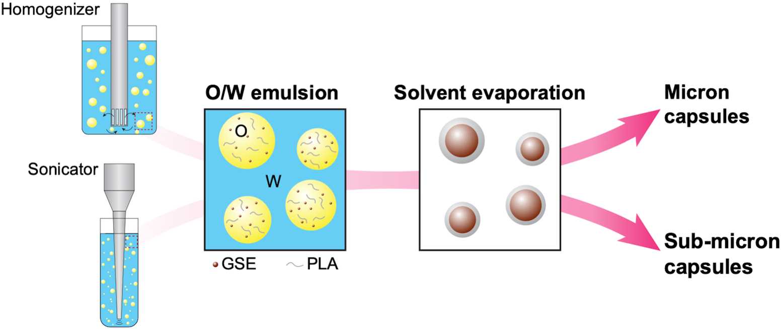
Methods: Sub-micron (SM) and micron (M) size capsules containing PACs were produced using a combination of emulsification and solvent evaporation techniques and characterized. Human dentin surfaces (n = 8) were etched (35% glycolic acid) and primed (15% enriched Vitis vinifera extract solution - VVe), followed by the application of an experimental adhesive containing 0 (control), 1.5 wt% of SM or M PAC-filled PLA capsules light cured for 40 s. A crown was built using commercial composite. After 24 h-immersion (37 °C) in simulated body fluid, specimens were serially sectioned into resin-dentin beams. Microtensile bond strength (TBS), micro-permeability and fracture pattern were assessed immediately and after 1 and 2 years. Data were statistically analyzed using two-way ANOVA and post-hoc test (α = 0.05).

Results: Polydisperse capsules were manufactured with average diameter of 0.36 µm and 1.08 µm for SM and M, respectively. The addition of capsules did not affect TBS (p = 0.889). After 2 years, TBS significantly decreased in SM (p = 0.006), whereas M showed similar initial values (p = 0.291). Overall, less micro-permeability was found in M than the control and SM group (p < 0.001). After 2 years, fractured surfaces from capsule-containing groups failed within the adhesive layer while control fractured at the bottom of the hybrid layer.

Significance: The addition of PAC-filled PLA microcapsules in a dental adhesive did not affect the bond strength while increased and sustained the protection against micro-permeability in the interface, likely due to release of PACs.

Keywords: Bioactive release; Bond strength; Polylactide capsules; Proanthocyanidins; Sustained release.


Figures

Figure 1.
Figure 1.
Schematic representation of the encapsulation technique
Figure 2.
Figure 2.
Size distribution and representative scanning electron microscopy images of sub-micron (A) and micron (B) capsules. The average diameter (Davg) for sub-micron and micron capsules were 0.36 μm and 1.08 μm, respectively. Images exposed polydisperse size capsules with smooth surfaces. Scale bar: 2 μm.
Figure 3.
Figure 3.
(A) Microtensile bond strength (in MPa, mean and standard deviation) and (B) Interfacial Micro-permeability (FEI- fluorescence emission intensity, mean values and standard error) of experimental groups as a function of time (24 h, 1 and 2 years). Different uppercase letters indicate statistically significant differences (p < 0.05) among time points within each experimental adhesive group. Same symbol indicates lack of statistical difference among adhesives within each time point (p > 0.05).
Figure 4.
Figure 4.
Fluorescence microscopy cross-sectional images (×40 magnification) of the resin-dentin interface after 24 h incubation. Open arrows indicate the presence of aggregates of sub-micron capsules. Close arrow illustrates isolated micron capsule located in the resin-adhesive interface. (A) control, (B) sub-micron, and (C) micron groups. Scale bar: 25 μm.
Figure 5.
Figure 5.
Representative scanning electron microscopy images of the resin-dentin interface after 2-year incubation at two magnifications, ×2,500 (left, scale bar: 10 μm) and ×5,000 (right, scale bar: 5 μm). Open arrows indicate the presence of capsules in the resin tags, observed in both sub-micron and micron groups. Closed arrows show small cracks initiating from the interface adhesive resin-capsules. Images in micron group evidenced degradation of the capsules’ shell wall and exposure of core. (A) control, (B) sub-micron, and (C) micron groups.
Figure 6.
Figure 6.
Representative scanning electron microscopy images of the debonded surface following microtensile test after 24 hours, 1 and 2 years. All images at ×3,000 magnification (scale bar: 5 μm). Asterisks indicate the presence of fractured resin tags within the dentin tubules, open arrows show crack propagation lines between capsule sites, and closed arrows reflect imprints of capsules that detached or degraded. (A) control, (B) sub-micron, and (C) micron groups.

References

    1. Demarco FF, Corrêa MB, Cenci MS, Moraes RR, Opdam NJM. Longevity of posterior composite restorations: not only a matter of materials. Dent Mater 2012;28:87–101. 10.1016/j.dental.2011.09.003. - DOI - PubMed
    1. Ástvaldsdóttir Á, Dagerhamn J, van Dijken JWV, Naimi-Akbar A, Sandborgh-Englund G, Tranæus S, Nilsson M. Longevity of posterior resin composite restorations in adults – A systematic review. J Dent 2015;43:934–54. 10.1016/j.jdent.2015.05.001. - DOI - PubMed
    1. Maas MS, Alania Y, Natale LC, Rodrigues MC, Watts DC, Braga RR. Trends in restorative composites research: what is in the future? Braz Oral Res 2017;31:e55. 10.1590/1807-3107BOR-2017.vol31.0055. - DOI - PubMed
    1. Zhou W, Liu S, Zhou X, Hannig M, Rupf S, Feng J, Peng X, Cheng L. Modifying adhesive materials to improve the longevity of resinous restorations. Int J Mol Sci 2019;20:723. 10.3390/ijms20030723. - DOI - PMC - PubMed
    1. Leme-Kraus AA, Aydin B, Vidal CMP, Phansalkar RM, Nam JW, McAlpine J, Pauli GF, Chen S, Bedran-Russo AK. Biostability of the proanthocyanidins-dentin complex and adhesion studies. J Dent Res 2017;96:406–12. 10.1177/0022034516680586. - DOI - PMC - PubMed

Publication types