Review
. 2022 Feb;36(2):294-302.
doi: 10.1038/s41433-021-01765-x. Epub 2022 Jan 9.

Complement cascade inhibition in geographic atrophy: a review

Affiliations
Review

Complement cascade inhibition in geographic atrophy: a review

Dhaval Desai et al. Eye (Lond). 2022 Feb.

Abstract

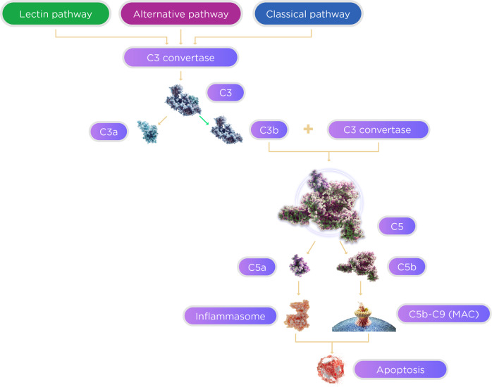
The pathophysiology of dry age-related macular degeneration (AMD) and specifically geographic atrophy (GA) has been linked to the complement cascade. This cascade is part of the innate immune system and is made up of the classical, alternative, and lectin pathways. The pathways comprise a system of plasma and membrane-associated serum proteins that are activated with identification of a nonself entity. A number of these proteins have been implicated in the development and progression of dry AMD. The three pathways converge at C3 and cascade down through C5, making both of these proteins viable targets for the treatment of dry AMD. In addition, there are a number of complement factors, CFB, CFD, CFH, and CFI, which are potential therapeutic targets as well. Several different complement-directed therapeutics are being studied for the treatment of dry AMD with the hope that one of these approaches will emerge as the first approved treatment for GA.

摘要: 干性年龄相关性黄斑变性 (AMD), 特别是地图样萎缩 (GA) 的病理生理学与补体级联有关。这种级联反应是先天免疫系统的一部分, 由经典, 替代和凝集素途径构成。这些通路包括一个与血浆和膜相关的血清蛋白系统, 该系统在识别非本体时被激活。其中的许多蛋白与干型AMD的发生和发展有关。这3条通路在C3处汇合, 并通过C5级联, 这两种蛋白为治疗干性AMD的可行靶点。此外, 还有一些补体因子, CFB, CFD, CFH和CFI, 也是潜在的治疗靶点。目前有几种不同的补体导向疗法也正在研究中, 以治疗干性AMD, 希望其中一种成为第一种被批准的GA治疗方法。.


Conflict of interest statement

Both authors are both employees of Iveric Bio.

Figures

Fig. 1
Fig. 1. The complement cascade.
The complement cascade is a highly conserved component of the innate immune system consisting of serum and membrane-bound proteins. This proteolytic cascade is activated through 3 pathways: the classical, alternative, and lectin. The activation pathways converge at C3 convertase, activation of which cleaves C3 into C3a and C3b subunits. As the cascade continues, C5 convertase cleaves C5, leading to inflammasome and MAC formation, ultimately resulting in apoptosis.
Fig. 2
Fig. 2. Evidence of complement activity in the formation of drusen [30].
Figure used with permission: Copyright (2005) National Academy of Sciences, USA.
Fig. 3
Fig. 3. Blocking C5a demonstrated a 60% reduction in retina loss in a mouse model.
Blocking C5 activation, which prevents the formation of both C5a and C5b, improved retinal protection over blocking C5a alone. [34]. C5a is required for photoreceptor loss. a Anti-C5a (a-C5a) inhibits C5a binding to C5aR on the surface of 293 cells transfected with C5aR (CD88). b Flow cytometry analysis of CD11b+CD45hi mononuclear phagocytes in the neural retina of mice treated with control or C5a neutralizing Abs 3 days following NaIO3 administration, n = 6. c Effect of C5a and C5 blocking Abs or isotype control Abs (Ctrl) on retina degeneration as measured by SD-OCT 7 days after NaIO3 administration. d Representative H&E stained sections of the central retina 7 days after NaIO3 administration. e POS length and RPE integrity measured in horizontal sections along the temporal-nasal axis of the mouse retina. Error bars indicate ±SEM, n = 3–4 naive and Ctrl, n = 7 a-C5a and a-C5. Naive = not NaIO3 treated. Scale bar = 10 μm. Central is 500 μm from optic nerve and periphery is 500–1750 μm. Experiments were repeated at least twice with similar results. *P < 0.05, **P < 0.01, ***P < 0.001, ****P < 0.0001 by one-way ANOVA with Tukey’s multiple comparisons test. INL inner nuclear layer, ONL outer nuclear layer, POS photoreceptor outer segments, RPE retinal pigmented epithelia, Ch choroid. Figure used under a Creative Commons Attribution 4.0 International License (http://creativecommons.org/licenses/by/4.0/; no changes were made).
Fig. 4
Fig. 4. Mean change from baseline in square root GA lesion area over 12 months.
AACP 2 mg B ACP 4 mg.

References

    1. Ferris FL, Wilkinson CP, Bird A, Chakravarthy U, Chew E, Csaky K, et al. Clinical classification of age-related macular degeneration. Ophthalmology. 2013;120:844–51. doi: 10.1016/j.ophtha.2012.10.036. - DOI - PMC - PubMed
    1. Jager RD, Mieler WF, Miller JW. Age-related macular degeneration. N Engl J Med. 2008;358:2606–17. doi: 10.1056/NEJMra0801537. - DOI - PubMed
    1. Al-Zamil W, Yassin S. Recent developments in age-related macular degeneration: a review. Clin Interv Aging. 2017;12:1313–30. doi: 10.2147/CIA.S143508. - DOI - PMC - PubMed
    1. De Jong PT. Elusive drusen and changing terminology of AMD. Eye. 2018;32:904–14. doi: 10.1038/eye.2017.298. - DOI - PMC - PubMed
    1. Davis MD, Gangnon RE, Lee LY, Hubbard LD, Klein BE, Klein R, et al. The Age-related Eye Disease Study severity scale for age-related macular degeneration: AREDS report no. 17. Arch Ophthalmol. 2005;123:1484–98. doi: 10.1001/archopht.123.11.1484. - DOI - PMC - PubMed

MeSH terms