

Compressive ultrasound can predict early pulmonary embolism onset in COVID patients
- PMID: 35000130
- PMCID: PMC8742694
- DOI: 10.1007/s40477-021-00625-4
Compressive ultrasound can predict early pulmonary embolism onset in COVID patients
Abstract
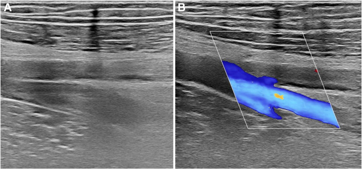
Purpose: To evaluate the usefulness of compressive ultrasound (CUS) for the diagnosis of deep vein thrombosis (DVT) in patients with SARS-CoV-2-related infection.
Methods: 112 hospitalized patients with confirmed SARS-CoV-2 infection were retrospectively enrolled. CUS was performed within 2 days of admission and consisted in the assessment of the proximal and distal deep venous systems. Lack of compressibility, or direct identification of an endoluminal thrombus, were the criteria used for the diagnosis of DVT. Pulmonary embolism (PE) events were investigated at computed tomography pulmonary angiography (CTPA) within 5 days of follow-up. Logistic binary regression was computed to determine which clinical and radiological parameters were independently associated with PE onset.
Results: Overall, the incidence of DVT in our cohort was about 43%. The most common district involved was the left lower limb (68.7%) in comparison with the right one (58.3%) while the upper limbs were less frequently involved (4.2% the right one and 2.1% the left one, respectively). On both sides, the distal tract of the popliteal vein was the most common involved (50% right side and 45.8% left side). The presence of DVT in the distal tract of the right popliteal vein (OR = 2.444 95%CIs 1.084-16.624, p = 0.038), in the distal tract of the left popliteal vein (OR = 4.201 95%CIs 1.484-11.885, p = 0.007), and D-dimer values (OR = 2.122 95%CIs 1.030-5.495, p = 0.003) were independently associated with the onset on PE within 5 days.
Conclusions: CUS should be considered a useful tool to discriminate which category of patients can develop PE within 5 days from admission.
Keywords: Coronavirus; Diagnostic imaging; Embolism and thrombosis; Infections; Thrombosis; Tomography, X-Ray computed; Ultrasonography.
© 2021. Società Italiana di Ultrasonologia in Medicina e Biologia (SIUMB).
Conflict of interest statement
Authors declared no conflict of interest.
Figures
Similar articles
-
Correlation between the site of pulmonary embolism and the extent of deep vein thrombosis: evaluation by computed tomography pulmonary angiography and computed tomography venography.Jpn J Radiol. 2011 Apr;29(3):171-6. doi: 10.1007/s11604-010-0533-y. Epub 2011 Apr 26. Jpn J Radiol. 2011. PMID: 21519990
-
A critical appraisal of non-invasive diagnosis and exclusion of deep vein thrombosis and pulmonary embolism in outpatients with suspected deep vein thrombosis or pulmonary embolism: how many tests do we need?Int Angiol. 2005 Mar;24(1):27-39. Int Angiol. 2005. PMID: 15876996 Review.
-
Chronic CT features in PE patients with co-existing DVT.Am J Emerg Med. 2021 Aug;46:126-131. doi: 10.1016/j.ajem.2021.03.031. Epub 2021 Mar 14. Am J Emerg Med. 2021. PMID: 33744749
-
Pulmonary Embolism and Deep Vein Thrombosis in COVID-19: A Systematic Review and Meta-Analysis.Radiology. 2021 Feb;298(2):E70-E80. doi: 10.1148/radiol.2020203557. Epub 2020 Dec 15. Radiology. 2021. PMID: 33320063 Free PMC article.
-
Deep vein thrombosis and pulmonary embolism among hospitalized coronavirus disease 2019-positive patients predicted for higher mortality and prolonged intensive care unit and hospital stays in a multisite healthcare system.J Vasc Surg Venous Lymphat Disord. 2021 Nov;9(6):1361-1370.e1. doi: 10.1016/j.jvsv.2021.03.009. Epub 2021 Apr 6. J Vasc Surg Venous Lymphat Disord. 2021. PMID: 33836287 Free PMC article.
Cited by
-
Ultrasound during the COVID-19 Pandemic: A Global Approach.J Clin Med. 2023 Jan 29;12(3):1057. doi: 10.3390/jcm12031057. J Clin Med. 2023. PMID: 36769702 Free PMC article. Review.
References
MeSH terms
LinkOut - more resources
Full Text Sources
Medical
Miscellaneous
