. 2022 Nov;76(5):1452-1465.
doi: 10.1002/hep.32324. Epub 2022 Apr 14.

Compromised hepatic mitochondrial fatty acid oxidation and reduced markers of mitochondrial turnover in human NAFLD

Affiliations

Compromised hepatic mitochondrial fatty acid oxidation and reduced markers of mitochondrial turnover in human NAFLD

Mary P Moore et al. Hepatology. 2022 Nov.

Abstract

Background and aims: NAFLD and its more-advanced form, steatohepatitis (NASH), is associated with obesity and is an independent risk factor for cardiovascular, liver-related, and all-cause mortality. Available human data examining hepatic mitochondrial fatty acid oxidation (FAO) and hepatic mitochondrial turnover in NAFLD and NASH are scant.

Approach and results: To investigate this relationship, liver biopsies were obtained from patients with obesity undergoing bariatric surgery and data clustered into four groups based on hepatic histopathological classification: Control (CTRL; no disease); NAFL (steatosis only); Borderline-NASH (steatosis with lobular inflammation or hepatocellular ballooning); and Definite-NASH (D-NASH; steatosis, lobular inflammation, and hepatocellular ballooning). Hepatic mitochondrial complete FAO to CO2 and the rate-limiting enzyme in β-oxidation (β-hydroxyacyl-CoA dehydrogenase activity) were reduced by ~40%-50% with D-NASH compared with CTRL. This corresponded with increased hepatic mitochondrial reactive oxygen species production, as well as dramatic reductions in markers of mitochondrial biogenesis, autophagy, mitophagy, fission, and fusion in NAFL and NASH.

Conclusions: These findings suggest that compromised hepatic FAO and mitochondrial turnover are intimately linked to increasing NAFLD severity in patients with obesity.


Conflict of interest statement

DECLARATION OF INTERESTS

No conflicts of interest, financial or otherwise, are declared by the authors.

Figures

Figure 1.
Figure 1.
Liver phenotype and measures of oxidative stress in humans. A) Representative liver H&E and B) trichrome staining, C) histological steatosis, inflammation, and ballooningNAFLD activity score, and D) histological fibrosis score. Values are presented as mean ± SE. CTRL = Control, NAFL = Nonalcoholic fatty liver, B-NASH = Borderline NASH, D-NASH = Definite-NASH. δ = main effect (P ≤ 0.05). *P ≤ 0.05, **P ≤ 0.01, ***P ≤ 0.001, ****P ≤ 0.0001.
Figure 2.
Figure 2.. Whole liver and isolated hepatic mitochondria ex-vivo fatty acid oxidation in humans.
Whole liver A) complete oxidation to CO2, incomplete, and total [1-14C] palmitate oxidation. B) Whole liver β-HAD activity and C) Whole liver complete palmitate oxidation across hepatic fibrosis scores. Isolated hepatic mitochondria D) complete oxidation to CO2, incomplete, and total [1-14C] palmitate oxidation. E) Isolated hepatic mitochondria β-HAD activity and F) Isolated hepatic mitochondria complete palmitate oxidation across hepatic fibrosis scores. Values are presented as mean ± SE. CTRL = Control, NAFL = Nonalcoholic fatty liver, B-NASH = Borderline NASH, D-NASH = Definite-NASH. δ = main effect (P ≤ 0.05), *P ≤ 0.05, **P ≤ 0.01, ***P ≤ 0.001, ****P ≤ 0.0001.
Figure 3.
Figure 3.. Measures of hepatic mitochondrial function, content and dynamics in humans with NAFL, B-NASH and D-NASH.
O2 consumption in isolated hepatic mitochondria A) malate + glutamate-stimulated, malate + palmitoylcarnitine-stimulated, and malate + octanoylcarnitine-stimulated; followed by the addition of adenosine diphosphate (ADP), succinate, and carbonyl cyanide 4-trifluoromethoxy-phenylhydrazone (FCCP). B) Palmitoyl-CoA stimulated H2O2 emission in isolated hepatic mitochondria across NAFLD and comparisons across histological inflammation and histological ballooning. C) Whole liver citrate synthase activity. D) Mitochondrial OXPHOS complexes protein content across hepatic fibrosis scores measured in whole liver, and representative Western blot images. E) Mitochondrial biogenesis markers mRNA expression in whole liver. F) Pgc1α mRNA expression across fibrosis scores. All Western blots were run on continuous gels. Values are presented as mean ± SE. CTRL = Control, NAFL = Nonalcoholic fatty liver, B-NASH = Borderline NASH, D-NASH = Definite-NASH. δ = main effect (P ≤ 0.05), P ≤ 0.05, **P ≤ 0.01, ***P ≤ 0.001, ****P ≤ 0.0001.
Figure 4.
Figure 4.. Mitochondrial structure and morphology with increasing NAFLD/NASH.
Representative transmission electron microscopy images of whole liver in patients CTRL, B-NASH, and D-NASH at A) 5,000x and B) 10,000x magnification.
Figure 5.
Figure 5.. Measures of autophagy and mitophagy in whole liver.
A) Protein content of autophagy markers in whole liver and representative Western blot images. B) LC3II/LC3I ratio across fibrosis scores and C) P62 protein content across fibrosis scores. D) BNIP3, PARKIN, and PINK1 protein content in whole liver and representative Western blot images. E) Bnip3 mRNA expression across fibrosis scores. F) Markers of mitochondrial fission and fusion protein content in whole liver and representative Western blot images. All Western blots were run on continuous gels. Values are presented as mean ± SE. CTRL = Control, NAFL = Nonalcoholic fatty liver, B-NASH = Borderline NASH, D-NASH = Definite-NASH. δ = main effect (P ≤ 0.05), *P ≤ 0.05, **P ≤ 0.01, ***P ≤ 0.001, ****P ≤ 0.0001.
Figure 6.
Figure 6.. Correlational Analysis
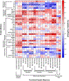
Correlation Heat map - showing Pearson’s correlation coefficients of functional ex vivo measures of hepatic fatty acid oxidation, O2 respiration, and ROS with protein content of markers related to mitochondrial content, quality control and oxidative stress. Protein content was measured via Western blot. Significant correlations are indicated on heat map with r value and significance level. Red indicates a positive correlation; blue indicates a negative correlation between measures. *P ≤ 0.05, **P ≤ 0.01, ***P ≤ 0.001, ****P ≤ 0.0001.
Figure 7.
Figure 7.. Ingenuity Pathway Analysis network revealing changes in the regulation of disease and biological functions for D-NASH versus CTRL.
Protein content of markers related to mitochondrial content, quality control and oxidative stress were measured via Western blot. Significant differences were determined using a one-way ANCOVA, with significance level set a P ≤ 0.05, and followed up with a Tukey post-hoc analysis. Values shown in Figure 7 are fold change and p-value of the individual proteins. CTRL = Control, D-NASH = Definite-NASH.

Comment in

References

    1. Chalasani N, Younossi Z, Lavine JE, Charlton M, Cusi K, Rinella M, Harrison SA, et al. The diagnosis and management of nonalcoholic fatty liver disease: Practice guidance from the American Association for the Study of Liver Diseases. Hepatology 2018;67:328–357. - PubMed
    1. Stepanova M, Rafiq N, Makhlouf H, Agrawal R, Kaur I, Younoszai Z, McCullough A, et al. Predictors of all-cause mortality and liver-related mortality in patients with non-alcoholic fatty liver disease (NAFLD). Digestive Diseases and Sciences 2013;58:3017–3023. - PubMed
    1. Laye MJ, Rector RS, Borengasser SJ, Naples SP, Uptergrove GM, Ibdah JA, Booth FW, et al. Cessation of daily wheel running differentially alters fat oxidation capacity in liver, muscle, and adipose tissue. Journal of Applied Physiology 2009;106:161–168. - PMC - PubMed
    1. Morris EM, McCoin CS, Allen JA, Gastecki ML, Koch LG, Britton SL, Fletcher JA, et al. Aerobic capacity mediates susceptibility for the transition from steatosis to steatohepatitis. The Journal of Physiology 2017;595:4909–4926. - PMC - PubMed
    1. Rector RS, Thyfault JP, Morris RT, Laye MJ, Borengasser SJ, Booth FW, Ibdah JA. Daily exercise increases hepatic fatty acid oxidation and prevents steatosis in Otsuka Long-Evans Tokushima Fatty rats. American Journal of Physiology - Gastrointestinal and Liver Physiology 2008;294:G619–626. - PubMed

Publication types