Review
. 2022 Jan 9;17(1):4.
doi: 10.1186/s13024-021-00506-8.

The role of inflammasomes in vascular cognitive impairment

Affiliations
Review

The role of inflammasomes in vascular cognitive impairment

Luting Poh et al. Mol Neurodegener. .

Abstract

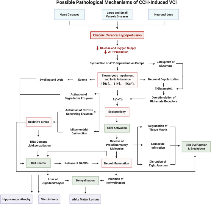
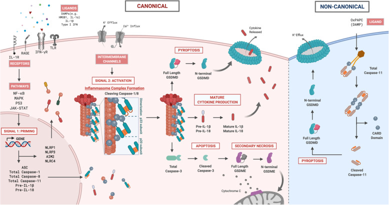
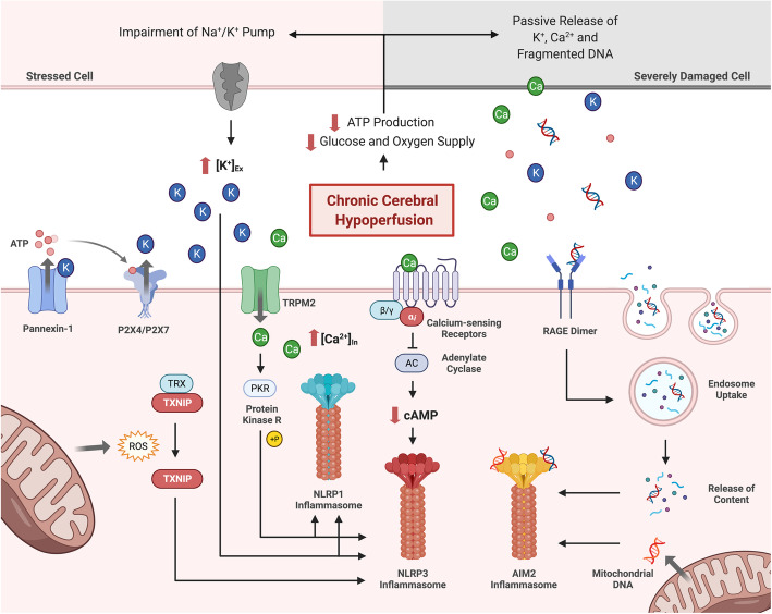
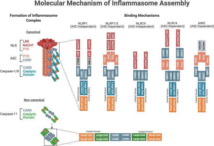
There is an increasing prevalence of Vascular Cognitive Impairment (VCI) worldwide, and several studies have suggested that Chronic Cerebral Hypoperfusion (CCH) plays a critical role in disease onset and progression. However, there is a limited understanding of the underlying pathophysiology of VCI, especially in relation to CCH. Neuroinflammation is a significant contributor in the progression of VCI as increased systemic levels of the proinflammatory cytokine interleukin-1β (IL-1β) has been extensively reported in VCI patients. Recently it has been established that CCH can activate the inflammasome signaling pathways, involving NLRP3 and AIM2 inflammasomes that critically regulate IL-1β production. Given that neuroinflammation is an early event in VCI, it is important that we understand its molecular and cellular mechanisms to enable development of disease-modifying treatments to reduce the structural brain damage and cognitive deficits that are observed clinically in the elderly. Hence, this review aims to provide a comprehensive insight into the molecular and cellular mechanisms involved in the pathogenesis of CCH-induced inflammasome signaling in VCI.

Keywords: Chronic cerebral Hypoperfusion; Inflammasome; Inflammation; Vascular cognitive impairment; Vascular dementia.


Conflict of interest statement

Not applicable.

Figures

Fig. 1
Fig. 1
A schematic diagram illustrating the possible pathological mechanisms of VCI. Cardiovascular disease is major contributor to early cerebral blood flow reduction in the disease progression of VCI. These conditions include heart disease (i.e. coronary artery disease and arrhythmias) that impairs the ejection of blood into the blood circulation; and small and large vessel diseases (i.e. atherosclerosis and arteriosclerosis), which narrow the vascular lumen and impede blood flow. Neuronal loss results in reduced production of angiogenesis regulators, leading to neurovascular unit uncoupling. These conditions converge to cause chronic cerebral hypoperfusion that reduces delivery of glucose and oxygen to the brain leading to decreased energy (i.e. ATP) production, resulting in bioenergetic impairment. Reduced ATP levels initiate a series of pathogenic molecular and cellular mechanisms. Firstly, the function of ATP-dependent transporters (i.e. Na+/K+ ATPase) are impaired leading to ionic imbalance (i.e. Na+ and Ca2+ influx, and K+ efflux) across the plasma membrane resulting in anoxic depolarization within neurons causing excitotoxicity. Moreover, increased levels of intracellular Ca2+ activate a wide variety of calcium-dependent ROS generating pathways in the mitochondria and cytosol contributing to oxidative stress. Finally, neuroinflammation is activated as stressed or injured cells release DAMPs that bind to PRRs to induce an inflammatory response. Under CCH, these molecular mechanisms influence each other within different cell types that result in the following pathogenic cellular mechanisms: glial activation, BBB dysfunction, cell death and demyelination. As pathogenic cellular mechanisms accumulate, they synergistically drive further damage eventually causing structural damage such as white matter lesions, microinfarcts and hippocampal atrophy. Each of these structural changes cause disruption to the neuronal network and functional connectivity that eventually leads to cognitive decline. Abbreviations: VCI, vascular cognitive impairment; ATP, adenosine triphosphate; ROS, reactive oxygen species; DAMPs, damage-associated molecular patterns; CCH, chronic cerebral hypoperfusion; BBB, blood brain barrier
Fig. 2
Fig. 2
A schematic diagram illustrating the canonical and non-canonical inflammasome signaling pathways in the brain during chronic cerebral hypoperfusion. In the canonical inflammasome pathway, two signals – priming and activation are involved. The first signal is the priming step whereby endogenous extracellular ligands (DAMPs) are able to bind onto its respective pattern recognition receptors (i.e. TLR, RAGE, IFN-γR, IL-1R) on neighbouring cells, activating several downstream regulatory pathways (i.e. NF-κB, MAPK, P53 and JAK-STAT), leading to increased gene expression of inflammasome components (i.e. receptors, adaptor and effector proteins) and both precursor IL-1β and IL-18 in the cytosol. Following priming, a second signal is required to activate the inflammasome receptor(s) to form a macromolecular platform that recruits the adaptor protein (i.e. ASC) and effector proteins (i.e. total caspase-1 and -8) to form a multi-protein complex termed an inflammasome. In the inflammasome complex, total caspase-1 and -8 undergo proximity-induced activation to form active cleaved caspase-1 and -8 that initiates several catalytic functions. First, cleaved caspase-1 and -8 induces mature cytokine production by cleaving precursors IL-1β and IL-18 into active mature IL-1β and IL-18 proinflammatory cytokines. Second, cleaved caspase-1 and -8 are able to initiate an inflammatory form of cell death by cleaving GSDMD-FL into GSDMD-NT. As more GSDMD-NTs are produced in the cytosol, these fragments self-oligomerize onto the plasma membrane to form a pore to facilitate the influx of water molecules to induce a lytic form of cell death known as pyroptosis. Third, cleaved caspase-1 and -8 can trigger apoptosis by cleaving total caspase-3 into active cleaved caspase-3. Moreover, active cleaved caspase-3 can also initiate another form of cell death known as secondary necrosis by cleaving GSDME-FL into GSDME-NT. Similar to GSDMD-NT, GSDME-NT can self-oligomerize to form pores on the plasma membrane; in addition to forming pores on the mitochondrial membrane, which results in cytochrome c release, further exacerbating apoptosis. In the non-canonical inflammasome pathway, total caspase-11 can be activated by binding to an endogenous ligand (i.e. OxPAPC) that allows oligomerization of total caspase-11. Such oligomerization initiates the proximity-induced activation of total caspase-11 to form active cleaved caspase-11. The non-canonical effector protein, cleaved caspase-11, can also directly cleave GSDMD-FL into GSDMD-NT to cause pore formation. It has been shown that K+ efflux resulting from pore formation can serve as an activation signal for canonical NLRP3 receptor activation, indicating cross-talk between the canonical and non-canonical inflammasome signalling pathways. Abbreviations: DAMPs, damage associated molecular patterns; HMGB1, high mobility group box protein 1; IL, interleukin; IFN, interferon; TLR, toll-like receptor; RAGE, receptor for advanced glycation end products; NF-κB, nuclear factor kappa-light-chain enhancer of activated B cells; MAPK, mitogen activated protein kinase; JAK/STAT, janus kinase-signal transducer and activator of transcription; Pre, precursor; GSDMD, gasdermin D; GSDME, gasdermin E
Fig. 3
Fig. 3
A schematic diagram illustrating the molecular structure of inflammasome receptor, adaptor and effector components. Members of the NLR family share similar structures: the NACHT domain is responsible for NLR oligomerization, while LRR is the inhibitory unit of the NACHT domain, keeping the receptor in its inactive state. The two other critical domains are the PYD domain and the caspase activation and recruitment domain (CARD) to facilitate interactions with other inflammasome components with similar domains to form the NLRP1, NLRP3 and NLRC4 inflammasome complex. Under the PYHIN family, the AIM2 inflammasome receptor has a PYD domain for adaptor binding and a HIN200 domain for dsDNA ligand binding. The adaptor protein ASC has both the PYD and CARD domain, serving as a linker protein between the inflammasome receptor and effector protein components. The three effector protein components share similar catalytic units (i.e. large and small units) and an N-terminal domain (i.e. CARD or DED) for inflammasome complex binding. Abbreviations: NLR, nucleotide-binding oligomerization domain-like receptor; NACHT, NAIP (neuronal apoptosis inhibitor protein) C2TA (class 2 transcription activator, of the MHC) HET-E (heterokaryon incompatibility) and TP1 (telomerase-associated protein 1); LRR, leucine-rich repeat; PYD, pyrin domain; CARD, caspase recruitment domain; NLRP1, NLR family pyrin domain containing 1; NLRP3, NLR family pyrin domain containing 3; NLRC4, NLR family CARD domain-containing protein 4; AIM2, absent in melanoma 2; HIN200, hematopoietic interferon-inducible nuclear proteins with a 200-amino-acid repeat; dsDNA, double-stranded DNA; ASC, apoptosis-associated speck-like protein containing a CARD; DED, death effector domain
Fig. 4
Fig. 4
A schematic diagram illustrating potential stimuli involved in inflammasome receptor activation during CCH. The precise molecular and cellular mechanisms of inflammasome receptor activation during CCH are unknown. However, relevant studies suggest several plausible mechanisms including - decreased intracellular K+ concentration, increased intracellular Ca2+ concentration, ROS production, DNA fragmentation and oxidized mitochondrial DNA. During CCH, lower cerebral blood flow reduces ATP production, and impairs ATP-dependent transporters such as the Na+/K+-ATPase pump, leading to K+ accumulation in the extracellular space. Alternatively, ATP released by damaged cells can bind to the P2X4 and P2X7 receptors on neighbouring cells, leading to the receptor opening and efflux of K+. In addition, damaged cells can also passively release K+ into the extracellular environment. Extracellular K+ can activate Pannexin-1 channels on the plasma membrane through a mechanism independent of the membrane potential. This further promotes the release of ATP into the extracellular space, creating a positive feedback loop for K+ efflux. Consequently, the accumulation of extracellular K+ and decrease in intracellular K+ levels can activate the NLRP3 receptor by inducing a conformational change that promotes oligomerization. During CCH, severely damaged necrotic cells may also release Ca2+ into the extracellular space, activating calcium-sensing receptors (CaSRs) on neighbouring cells. Activated CaSRs inhibit the activity of adenylate cyclase, reducing the conversion of ATP to cAMP. As cAMP is an inhibitor for NLRP3, a reduction in cAMP levels in the cytosol can promote NLRP3 inflammasome activity. Ca2+ can also promote inflammasome activation through the TRPM2 Ca2+ channel during CCH. As Ca2+ enters the cell via the TRPM2 channel, it enables protein kinase R (PKR) in the cytoplasm to phosphorylate NLRP1 and NLRP3 receptors resulting in inflammasome activation. CCH also caused a substantial degree of oxidative stress and the production of ROS in the cell. ROS can interact with the TXNIP-TRX complex to release TXNIP from TRX, allowing it to bind to the NLRP3 receptor for subsequent inflammasome activation. CCH induces AIM2 inflammasome activation via the production and release of fragmented dsDNA. Severely damaged cells and mitochondria are the source of fragmented dsDNA during CCH. While intracellular mitochondrial dsDNA interacts directly with the AIM2 receptor in the cytosol, extracellular dsDNA enters the cell via the facilitation of RAGE. When RAGE detects the presence of dsDNA in extracellular space, it promotes endosomal DNA uptake of the cell. The dsDNA will then bind onto the HIN-domain of the AIM2 receptor, releasing the receptor from its autoinhibitory state. This allows the AIM2 receptor to oligomerize and initiate inflammasome activation. Abbreviations: CCH, chronic cerebral hypoperfusion; ROS, reactive oxygen species; ATP, adenosine triphosphate; P2X4, P2X purinoceptor 4; P2X7, P2X purinoceptor 7; NLR, nucleotide-binding oligomerization domain-like receptor; NLRP1, NLR family pyrin domain containing 1; NLRP3, NLR family pyrin domain containing 3; cAMP, cyclic adenosine monophosphate; TRPM2, transient receptor potential melastatin 2; TXNIP, thioredoxin-interacting protein; TRX, thioredoxin; AIM2, absent in melanoma 2; HIN200, hematopoietic interferon-inducible nuclear proteins; dsDNA, double-stranded DNA; RAGE, receptor for advanced glycation end-products
Fig. 5
Fig. 5
A schematic diagram illustrating the assembly of the canonical and non-canonical inflammasome complexes. The formation of the canonical inflammasome complex requires the activation of inflammasome receptors from the second signal. As the LRR inhibitory unit unfolds from the NACHT domain, the receptors are in an “open” structure for homotypic oligomerization through their NACHT domain. Subsequently, the PYD domain recruits the adaptor protein via the PYD domain on ASC. As numerous ASC adaptor proteins comes together, they will form a filamentous structure with their CARD domain exposed. Consequently, effector proteins with their CARD domain can bind to the filamentous structure via CARD-CARD interactions. Such protein aggregation triggers proximity-induced activation of total caspase-1, and − 8, leading to cleavage of the inter-domain linker between the large and small units, producing active cleaved caspase-1 and -8. The above mentioned ASC-dependent binding mechanism generally applies to all inflammasome complexes. However, NLRP1 can form an inflammasome complex without the adaptor ASC. Using its C-terminal CARD domain, NLRP1 binds to the effector protein via the CARD-CARD domain. NLRC4 can also adopt the same ASC-independent binding mechanisms with the CARD domain on the receptor. In the non-canonical inflammasome pathway, caspase-11 can undergo homo-oligomerization in the absence of a receptor and adaptor protein component via the CARD-CARD domain interaction. Similar to the activation of total caspase-1 and -8, cleaved caspase-11 is produced upon proximity-induced activation. Abbreviations: NLR, nucleotide-binding oligomerization domain-like receptor; NACHT, NAIP (neuronal apoptosis inhibitor protein) C2TA (class 2 transcription activator, of the MHC) HET-E (heterokaryon incompatibility) and TP1 (telomerase-associated protein 1); LRR, leucine-rich repeat; PYD, pyrin domain; CARD, caspase recruitment domain; NLRP1, NLR family pyrin domain containing 1; NLRP3, NLR family pyrin domain containing 3; NLRC4, NLR family CARD domain-containing protein 4; AIM2, absent in melanoma 2; HIN200, hematopoietic interferon-inducible nuclear proteins with a 200-amino-acid repeat; dsDNA, double-stranded DNA; ASC, apoptosis-associated speck-like protein containing a CARD; DED, death effector domain

References

    1. World Health Organization . Alzheimer’s disease international. Dementia: a public health priority. 2012.
    1. Prince M, Bryce R, Albanese E, Wimo A, Ribeiro W, Ferri CP. The global prevalence of dementia: a systematic review and metaanalysis. Alzheimers Dement. 2013;9(1):63–75.e2. - PubMed
    1. Lobo A, Launer LJ, Fratiglioni L, Andersen K, Di Carlo A, Breteler MMB, et al. Prevalence of dementia and major subtypes in Europe: a collaborative study of population-based cohorts. Neurology. 2000;54(11 SUPPL. 5):S4–S9. - PubMed
    1. Di Carlo A, Baldereschi M, Amaducci L, Lepore V, Bracco L, Maggi S, et al. Incidence of dementia, Alzheimer’s disease, and vascular dementia in Italy. The ILSA study. J Am Geriatr Soc. 2002;50(1):41–48. - PubMed
    1. Fujihara S, Brucki SMD, Rocha MSG, Carvalho AA, Piccolo AC. Prevalence of presenile dementia in a tertiary outpatient clinic. Arq Neuropsiquiatr. 2004;62(3 A):592–595. - PubMed

Publication types