Review
. 2022 Jan 9;13(1):3.
doi: 10.1186/s13244-021-01138-8.

Active pulmonary tuberculosis: something old, something new, something borrowed, something blue

Affiliations
Review

Active pulmonary tuberculosis: something old, something new, something borrowed, something blue

Maria T A Wetscherek et al. Insights Imaging. .

Abstract

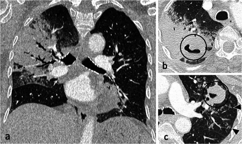
Tuberculosis remains a major global health issue affecting all countries and age groups. Radiology plays a crucial role in the diagnosis and management of pulmonary tuberculosis (PTB). This review aims to improve understanding and diagnostic value of imaging in PTB. We present the old, well-established findings ranging from primary TB to the common appearances of post-primary TB, including dissemination with tree-in-bud nodularity, haematogenous dissemination with miliary nodules and lymphatic dissemination. We discuss new concepts in active PTB with special focus on imaging findings in immunocompromised individuals. We illustrate PTB appearances borrowed from other diseases in which the signs were initially described: the reversed halo sign, the galaxy sign and the cluster sign. There are several radiological signs that have been shown to correlate with positive or negative sputum smears, and radiologists should be aware of these signs as they play an important role in guiding the need for isolation and empirical anti-tuberculous therapy.

Keywords: Computed tomography; Imaging; Immunocompromised; Pulmonary tuberculosis; X-ray.


Conflict of interest statement

The authors declare that they have no competing intersts.

Figures

Fig. 1
Fig. 1
A 37-year-old female had a routine chest radiograph (a) which demonstrated a solitary pulmonary nodule in the left mid zone (arrow). CT (lung window, axial plane—(b) confirms the presence of a 27 mm nodule in the lingula (arrow) with adjacent tiny satellite nodules (arrowheads) but no lymphadenopathy. Sputum smears were negative but TB culture from bronchoalveolar lavage was positive for M. Tuberculosis
Fig. 2
Fig. 2
An 83-year-old male presented with pyrexia of unknown origin. CT (lung window) demonstrates extensive consolidation in the right upper lobe (coronal plane—a), bilateral thick-walled cavities (axial planes—b, c, circled) and centrilobular nodules with a tree-in-bud appearance (arrowheads)
Fig. 3
Fig. 3
A 41-year-old male presented with chronic cough, weight loss and fevers. CT (lung window) demonstrates bilateral areas of centrilobular and tree-in-bud nodules (coronal plane—a, circled; axial plane—c) and consolidation in the right upper lobe (axial plane—b). There are also cavitating lesions with thick walls (arrow, a)
Fig. 4
Fig. 4
A 35-year-old male presented with a 4-week history of productive cough, anorexia and weight loss. Initial CT demonstrates tracheal and left main bronchus wall thickening with irregularity suggestive of ulcerations (lung window, coronal plane—a, arrow). There is also a thick-walled cavity with surrounding nodularity (arrowheads). The appearances significantly improved following TB treatment as demonstrated on a follow-up CT performed two months later (lung window, coronal plane—b)
Fig. 5
Fig. 5
A 44-year-old female presented with symptoms of lethargy, loss of appetite, vomiting and fevers. She had been diagnosed with HIV two months prior but was not compliant with antiretroviral therapy. Her CD4 count at the time of presentation was 110 cells/mm3. CT demonstrates miliary nodules (lung window, coronal plane, maximum intensity projection—a) and an enlarged right paratracheal node with central low attenuation (mediastinal window, coronal plane—b, arrow) consistent with necrosis
Fig. 6
Fig. 6
A 34-year-old male with Crohn’s disease presented with recurrent pyrexia and raised inflammatory markers. CT demonstrates a 29 mm subcarinal node with central low attenuation (mediastinal window, axial plane—a, arrow) invading into the right main bronchus (lung window, coronal plane—b, arrow). There is also right lower lobe consolidation and a small right pleural effusion (lung widow, axial plane—c). CT virtual bronchoscopy demonstrates the node protruding into the lumen of the right main bronchus (d) with corresponding bronchoscopy image (e). The patient underwent cryoprobe removal of the lymph node tissue successfully (bronchoscopy image—f)
Fig. 7
Fig. 7
A 29-year-old male presented with cough and haemoptysis. CT (lung window, coronal planes—a, b) demonstrates a cluster of tiny nodules within the apicoposterior segment of the left upper lobe with perilymphatic distribution
Fig. 8
Fig. 8
A 37-year-old male presented with intermittent pyrexia, sweats, rigors, reduced appetite and weight loss. Post-contrast CT shows a moderate volume left pleural effusion with pleural thickening and enhancement (mediastinal window, axial plane—a, arrowheads). There are also multifocal areas of perilymphatic nodularity (lung window, axial plane—b) and bilateral paratracheal and mediastinal lymphadenopathy (arrows, a). Pleural biopsy confirmed the presence of acid-fast bacilli
Fig. 9
Fig. 9
A 47-year-old female with a known diagnosis of HIV presented with weight loss, night sweats and worsening dyspnoea. Her CD4 count at presentation was 120 cells/mm3. CT (lung window) demonstrates thick-walled irregular cavities (coronal plane—a, arrow), extensive bilateral upper lobe centrilobular and tree-in-bud nodules and nodular reversed halo sign in the apical segment of the right lower lobe (axial plane—c, arrowhead)
Fig. 10
Fig. 10
A 74-year-old male with HIV and a CD4 count of 10 cells/mm3 was started on antiretroviral therapy. He developed immune reconstitution inflammatory syndrome with worsening imaging findings in keeping with paradoxical reaction. Compared to the CT from three months prior (mediastinal window, axial plane—a), there are enlarged centrally necrotic precarinal lymph nodes (mediastinal window, axial plane—b, arrow) and necrotic nodules in the right upper lobe (arrowheads). These were avid on PET-CT (c)
Fig. 11
Fig. 11
A 37-year-old male with active pulmonary TB presented with a 6-month history of weight loss and night sweats. CT (lung window) demonstrates nodular reversed halo sign in the right lower and middle lobe (axial plane—a, coronal plane—c), and cavitating lesions in the left upper lobe (coronal plane—b, circled). Image (d) is of a 25-year-old male with cryptogenic organising pneumonia who presented with cough and night sweats. CT (lung window, coronal plane) demonstrates multiple areas of rim consolidation reversed halo sign
Fig. 12
Fig. 12
A 27-year-old male with Crohn’s disease on treatment with infliximab presented with a cough that was not resolving despite antibiotic therapy. CT demonstrates extensive centrilobular and tree-in-bud nodules with a mid-upper zone predominance (lung window, coronal plane—a). There are also small volume mediastinal and hilar lymph nodes (mediastinal window, axial plane—b). Ziehl–Neelsen stain was positive for AFB (c)

References

    1. World Health Organization (2020) Global tuberculosis report 2020, Geneva. Available via https://apps.who.int/iris/bitstream/handle/10665/336069/9789240013131-en... Accessed 24 April 2021
    1. Rozenshtein A, Hao F, Starc MT, Pearson GDN. Radiographic appearance of pulmonary tuberculosis: dogma disproved. AJR Am J Roentgenol. 2015;204:974–978. doi: 10.2214/AJR.14.13483. - DOI - PubMed
    1. Marais BJ, Parker SK, Verver S, Warren RM (2009) Primary and postprimary or reactivation tuberculosis: time to revise confusing terminology? AJR Am J Roentgenol 192:W198; author reply W199–200. 10.2214/AJR.08.1726 - PubMed
    1. Nachiappan AC, Rahbar K, Shi X (2017) Pulmonary tuberculosis: role of radiology in diagnosis and management. Radiographics 37:52–72. 10.1148/rg.2017160032 - PubMed
    1. Burrill J, Williams CJ, Bain G, Conder G, Hine AL, Misra RR (2007) Tuberculosis: a radiologic review. Radiographics 27:1255–1273. 10.1148/rg.275065176 - PubMed

LinkOut - more resources