Review
. 2022 Sep;32(5):e13052.
doi: 10.1111/bpa.13052. Epub 2022 Jan 9.

Variable histopathology features of neuronal dyslamination in the cerebral neocortex adjacent to epilepsy-associated vascular malformations suggest complex pathogenesis of focal cortical dysplasia ILAE type IIIc

Affiliations
Review

Variable histopathology features of neuronal dyslamination in the cerebral neocortex adjacent to epilepsy-associated vascular malformations suggest complex pathogenesis of focal cortical dysplasia ILAE type IIIc

Hajime Miyata et al. Brain Pathol. 2022 Sep.

Abstract

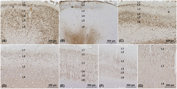
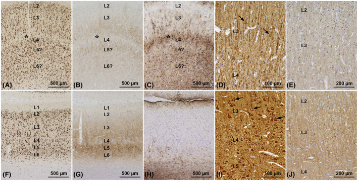
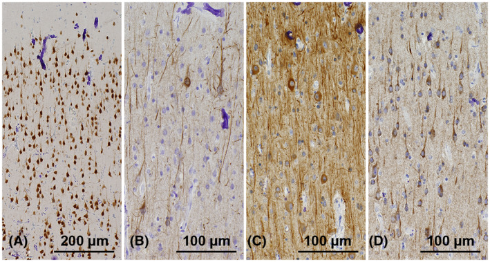
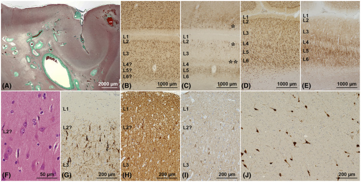
Focal cortical dysplasia type IIIc (FCD-IIIc) is histopathologically defined by the International League Against Epilepsy's classification scheme as abnormal cortical organization adjacent to epilepsy-associated vascular malformations (VM). However, the incidence of FCD-IIIc, its pathogenesis, or association with the epileptogenic condition remains to be clarified. We reviewed a retrospective series of surgical brain specimens from 14 epilepsy patients with leptomeningeal angiomatosis of Sturge-Weber syndrome (LMA-SWS; n = 6), cerebral cavernous malformations (CCM; n = 7), and an arteriovenous malformation (AVM; n = 1) to assess the histopathological spectrum of FCD-IIIc patterns in VM. FCD-IIIc was observed in all cases of LMA-SWS and was designated as cortical pseudolaminar sclerosis (CPLS). CPLS showed a common pattern of horizontally organized layer abnormalities, including neuronal cell loss and astrogliosis, either manifesting predominantly in cortical layer (L) 3 extending variably to deeper areas with or without further extension to L2 and/or L4. Another pattern was more localized, targeting mainly L4 with extension to L3 and/or L5. Abnormal cortical layering characterized by a fusion of L2 and L3 or L4-L6 was also noted in two LMA-SWS cases and the AVM case. No horizontal or vertical lamination abnormalities were observed in the specimens adjacent to the CCM, despite the presence of vascular congestion and dilated parenchymal veins in all VM. These findings suggest that FCD-IIIc depends on the type of the VM and developmental timing. We further conclude that FCD-IIIc represents a secondary lesion acquired during pre- and/or perinatal development rather than following a pathomechanism independent of LMA-SWS. Further studies will be necessary to address the selective vulnerability of the developing cerebral neocortex in LMA-SWS, including genetic, encephaloclastic, hemodynamic, or metabolic events.

Keywords: cerebral neocortex; epilepsy surgery; focal cortical dysplasia; neuron; pathogenesis; vascular malformation.


Conflict of interest statement

The authors declare that they have no conflicts of interest.

Figures

FIGURE 1
FIGURE 1
Leptomeningeal angiomatosis and cortical calcifications in SWS. (A–C) This surgical specimen was obtained from a 44‐year‐old male epilepsy patient with SWS (case 1 in Table 1). (A) A collection of dilated and congestive venous vessels in the subarachnoid space, i.e., leptomeningeal angiomatosis (LMA), and typical cortical calcifications of tram‐track pattern mainly affecting layer (L) 2 and L4 (asterisks). LFB‐HE staining. (B) Parenchymal veins in the white matter (arrows) are also significantly dilated and congested. Calcifications are accentuated in the superficial cortical area (asterisk). Elastica‐Masson staining. (C) Conspicuous string‐like, vertically oriented linear calcifications along the microvessels are accentuated in L2–L4. Larger plaque‐like calcifications are also accumulating mainly in L4 (asterisk). Immunohistochemistry for NeuN with hematoxylin counterstain. These features suggest the underlying hemodynamic abnormalities in the brain parenchyma and cortical areas where lesions are more likely to occur in patients with SWS
FIGURE 2
FIGURE 2
FCD‐IIIc with CPLS in superficial cortical areas. (A–D) The resection specimen was obtained from a 6‐year‐old epileptic male with SWS (case 6 in Table 1). (A) A horizontal zone of neuronal loss involving the deeper layer (L) 2 and superficial L3 (asterisk). Immunohistochemistry for NeuN with hematoxylin counterstain. (B) The lesion shown in panel A is accompanied by GFAP‐immunoreactive astrogliosis and termed, therefore, cortical pseudolaminar sclerosis (CPLS). This lesion pattern is reminiscent of temporal lobe sclerosis described in epilepsy patients with hippocampal sclerosis. (C) A more extensive lesion affecting the entire L3, extending partially to L2 and L4 (asterisk). NeuN immunohistochemistry. (D) The remaining neurons in L2 and L3 are immunoreactive for MAP1b. (E–G) The resection specimen from case 1. (E) NeuN immunohistochemistry shows a normal 6‐layered neuronal arrangement in the parietal cortex. One may argue that the microcolumnar arrangement of neurons in particularly thick L3 represents a structural abnormality; however, this is a standard feature for the normal parietal cortex. (F) In addition, immunohistochemistry for non‐phosphorylated neurofilament H (NF‐H) in the same area in panel E demonstrates NF‐H positive neurons in L3, L5, and L6, also indicating a normally developed layered cortical organization. (G) A magnified view of the area including L2–L4 in panels E and F demonstrating MAP1b immunoreactive neurons in L2 to shallower L3
FIGURE 3
FIGURE 3
FCD‐IIIc with CPLS in deeper cortical areas. The resection specimen from a 2.5‐year‐old epileptic male with SWS (case 2 in Table 1) showing FCD‐IIIc involving layer (L) 3 to L6 with CPLS in the deeper cortical areas (A–E) compared to normal‐appearing, although thin, hexalaminar neocortex adjacent to this FCD‐IIIc on the same specimen (F–J). (A) Neuronal cell loss is evident, affecting the putative deepest L3 and shallower L4 (asterisk). In addition, the separations between L3 and L4 and between L4, L5, and L6 are uncertain. For comparison, see panel F taken at the same magnification from the adjacent cortex of the same specimen showing a structurally normal‐appearing neocortex. NeuN immunohistochemistry with hematoxylin counterstain. (B) Faint NF‐H expression is observed in deeper L3, L5, and L6, and the border between L3 and L4 is uncertain because of the CPLS (asterisk). For comparison, see panel G showing stronger NF‐H immunoreactivities in L3, L5, and L6. (C) The pseudolaminar neuronal loss shown in panels A and B is accompanied by significant GFAP‐positive astrogliosis (asterisk). For comparison, see panel H showing Chaslin's gliosis in L1 only. (D) MAP2‐positive hypertrophic pyramidal neurons are scattered in L3 (arrows). By contrast, most neurons in the structurally normal‐appearing neocortex are more strongly immunoreactive for MAP2 in their cytoplasm, as shown in panel I, and neurons in L3 are larger than those in L5, a feature characterizing hypertrophic neurons (arrows in I). Panels D and I represent magnified views of L3 and L4 shown in panels A–C and L3–L5 shown in panels F–H, respectively. (E) Most pyramidal neurons in L3 are weakly immunoreactive for MAP1b in FCD‐IIIc than the adjacent neocortex shown in panel J. Panels E and J represent magnified views of L2 and L3 shown in panels A–C and L2–L4 shown in panels F–H, respectively
FIGURE 4
FIGURE 4
Histological and immunohistochemical findings on iron metabolism. (A–C) The resection specimen obtained from a 2‐year‐old epileptic male with LMA‐SWS (case 6 in Table 1). (D–F) The resection specimen obtained from a 2.5‐year‐old epileptic male with LMA‐SWS (case 2 in Table 1). (A, D) Lesions of cortical pseudolaminar sclerosis (CPLS) in the superficial and deeper cortex are accompanied by the accumulation of HLA‐DP, DQ, DR‐positive activated microglial cells of varying density. (B, E) No obvious Fe3+ depositions by Prussian blue staining are observed throughout the specimens examined, including the CPLS. (C, F) Ferritin expression in the cortex with CPLS is confined to a small number of oligodendrocytes. (G–I) The resection specimen obtained from a 37‐year‐old epileptic female with CCM (case 13 in Table 1). (G) Prussian blue staining in the cerebral cortex with hemosiderin depositions adjacent to CCM demonstrating the presence of Fe3+ in reactive astrocytes (arrows), microglial cells (arrowheads), and a proportion of neurons (double arrowheads). (B) Immunohistochemistry for ferritin heavy chain in the same area in panel A demonstrating immunoreactivities in reactive astrocytes (arrows), microglial cells (arrowheads), and a proportion of neurons (double arrowheads). (C) Ferritin expression in the Prussian blue‐defined Fe3+‐free area remote from the vascular malformations is confined to oligodendrocytes in the cerebral cortex and white matter. Scale bars in all panels indicate 100 µm
FIGURE 5
FIGURE 5
Abnormal organization of cortical layers L2 and L3 even without CPLS in SWS. The cerebral cortex without CPLS taken from case 1 (A–D, Table 1, and Figure 1). (A) Only a few neurons can be recognized as granular, whereas many show a pyramidal shape. In addition, the border between layer (L) 2 and L3 is blurred. L1 is presented at the upper margin. NeuN immunohistochemistry with hematoxylin counterstain. Panels B–D represent magnified views from the same region shown in panel A, including the presumed border between L2 and shallower L3. Some pyramidal neurons show accumulation of NF‐H (B) and/or MAP2 (C) in their cytoplasm. (D) Many pyramidal neurons are immunoreactive for MAP1b
FIGURE 6
FIGURE 6
FCD‐IIIc associated with AVM. (A) The resection specimen was obtained from a 26‐year‐old male epilepsy patient with AVM (case 14 in Table 1) showing a collection of irregularly dilated malformed vessels located mainly in the subcortical white matter, i.e., a nidus. Elastica‐Masson staining. (B) In the cortex adjacent to the AVM, the borders between layers (L) 2 to L6 are indistinct and blurred, compatible with FCD‐IIIc. NeuN immunohistochemistry with hematoxylin counterstain. (C) Immunohistochemistry for non‐phosphorylated neurofilament H (NF‐H) in the same area shown in panel B, demonstrating overall weak expression of NF‐H (compared to panel E) with few neurons accumulating NF‐H in L2 to shallower L3 (single asterisks), deep L3 (double asterisks), and L5, suggesting a preexisting hexalaminar organization of the neocortex. (D) NeuN immunohistochemistry showing a normal 6‐layered neuronal arrangement in the cerebral cortex in the same specimen but remote from the AVM, compared to panel B. (E) NF‐H expression is accentuated in L3, L5, and L6 in the same area shown in panel D. Of note, the FCD‐IIIc cortex presented in panels B, C is thinner than the structurally normal‐appearing cortex shown in panels D, E. (F) A magnified view of L2 within FCD‐IIIc, presented in panels B and C, demonstrating large pyramidal neurons, loss of neuronal polarity, and lack of granular neurons. LFB‐HE staining. (G) A magnified view of the area including L1 to L3, shown in panels B and C, demonstrating large pyramidal neurons with strong immunoreactivity for NF‐H, loss of neuronal polarity, and dystrophic neurites in L2 and shallower L3. (H) Large pyramidal neurons with strong immunoreactivity for MAP2 observed in the same area presented in panel G. (I) Large pyramidal neurons with immunoreactivity for MAP1b observed in the same area shown in panels G and H, and consistent with hypertrophic neurons. (J) Abundant heterotopic neurons are observed in the subcortical white matter in between malformed vessels

Similar articles

Cited by

References

    1. Challa VR. Vascular malformations and angiomas. In: Kalimo H, editor. Pathology & genetics. Cerebrovascular diseases. Basel: ISN Neuropath Press. 2005; p. 119–24.
    1. Blumcke I, Spreafico R, Haaker G, Coras R, Kobow K, Bien CG, et al. Histopathological findings in brain tissue obtained during epilepsy surgery. N Eng J Med. 2017;377:1648–56. - PubMed
    1. Rosenow F, Alonso‐Vanegas MA, Baumgartner C, Blümcke I, Carreño M, Gizewski ER, et al. Cavernoma‐related epilepsy: review and recommendations for management – report of the Surgical Task Force of the ILAE Commission on Therapeutic Strategies. Epilepsia. 2013;54:2025–35. - PubMed
    1. Ivens S, Kaufer D, Flores LP, Bechmann I, Zumsteg D, Tomkins O, et al. TGF‐β receptor‐mediated albumin uptake into astrocytes is involved in neocortical epileptogenesis. Brain. 2007;130:535–47. - PubMed
    1. Raabe A, Schmitz AK, Pernhorst K, Grote A, von der Brelie C, Urbach H, et al. Cliniconeuropathologic correlations show astroglial albumin storage as a common factor in epileptogenic vascular lesions. Epilepsia. 2012;53:539–48. - PMC - PubMed

Publication types

MeSH terms