

3-hydroxy butyrate dehydrogenase 2 deficiency aggravates systemic lupus erythematosus progression in a mouse model by promoting CD40 ligand demethylation
- PMID: 35001849
- PMCID: PMC8973909
- DOI: 10.1080/21655979.2022.2025694
3-hydroxy butyrate dehydrogenase 2 deficiency aggravates systemic lupus erythematosus progression in a mouse model by promoting CD40 ligand demethylation
Abstract
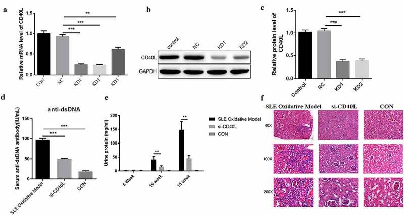
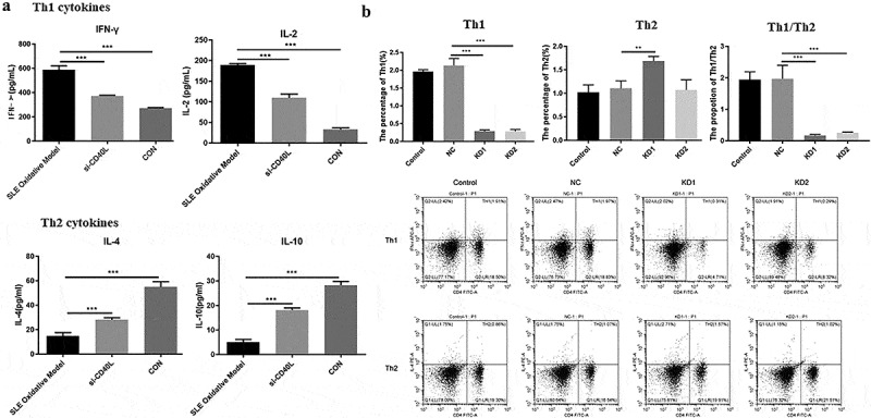
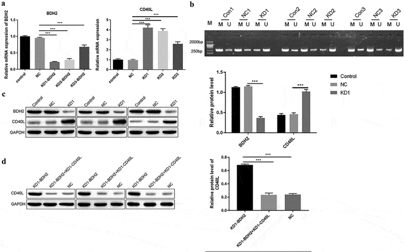
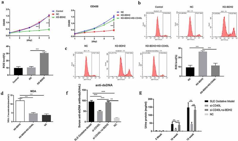
The implications of the CD40-CD40 ligand (CD40L) signaling pathway in systemic lupus erythematosus (SLE) were well documented, due to its important role among immune cells. Previous research found that 3-hydroxy butyrate dehydrogenase 2 (BDH2), a modulator of intracellular iron homeostasis and iron transportation promoted the pathogenic process of SLE by regulating the demethylation of cd70, cd11a, and cd40l genes among CD4 + T cells. The purpose of this study was to explore the role of BDH2 in oxidative damage-induced SLE. First, CD4 + T cells treated with H2O2 were injected into the tail vein of mice to establish a lupus model. CD40L knockdown significantly decreased CD40L expression on CD4 + T cells in the spleen of SLE mice. Compared with SLE model mice, the levels of serum anti-dsDNA antibody and urinary protein in the CD40L interference group were significantly decreased. CD40L knockdown alleviated the immune complex glomerulonephritis in syngeneic SLE mice. Moreover, the levels of IFN-γ and IL-2 were decreased. However, IL-4 and IL-10 levels were significantly upregulated in the serum of CD40L knockdown SLE mice, compared with SLE model mice. Accordingly, CD40L knockdown reduced Th1/Th2 percentage in SLE mice. Inhibiting the expression of BDH2 of CD4 + T cells promoted the demethylation of CD40L, while it inhibited cell proliferation, elevated oxidative stress through increased expression of CD40L, and thus, promoted the progress of SLE. Our results demonstrate that BDH2 aggravates the pathologic progression of SLE in mice, by increasing the demethylation level of CD40L among CD4 + T cells.
Keywords: 3-hydroxy butyrate dehydrogenase 2; CD4+ T cells; CD40L; Systemic lupus erythematosus; demethylation.
Conflict of interest statement
No potential conflict of interest was reported by the author(s).
Figures
Similar articles
-
Downregulation of BDH2 modulates iron homeostasis and promotes DNA demethylation in CD4+ T cells of systemic lupus erythematosus.Clin Immunol. 2018 Feb;187:113-121. doi: 10.1016/j.clim.2017.11.002. Epub 2017 Nov 4. Clin Immunol. 2018. PMID: 29113828
-
Roles of 1,25(OH)2D3 and Vitamin D Receptor in the Pathogenesis of Rheumatoid Arthritis and Systemic Lupus Erythematosus by Regulating the Activation of CD4+ T Cells and the PKCδ/ERK Signaling Pathway.Cell Physiol Biochem. 2016;40(3-4):743-756. doi: 10.1159/000453135. Epub 2016 Dec 5. Cell Physiol Biochem. 2016. PMID: 27915349
-
CD4(+) T cells epigenetically modified by oxidative stress cause lupus-like autoimmunity in mice.J Autoimmun. 2015 Aug;62:75-80. doi: 10.1016/j.jaut.2015.06.004. Epub 2015 Jul 9. J Autoimmun. 2015. PMID: 26165613 Free PMC article.
-
Phoenix from the flames: Rediscovering the role of the CD40-CD40L pathway in systemic lupus erythematosus and lupus nephritis.Autoimmun Rev. 2020 Nov;19(11):102668. doi: 10.1016/j.autrev.2020.102668. Epub 2020 Sep 14. Autoimmun Rev. 2020. PMID: 32942031 Review.
-
Regulation of CD40 ligand expression in systemic lupus erythematosus.Curr Opin Rheumatol. 2001 Sep;13(5):361-9. doi: 10.1097/00002281-200109000-00004. Curr Opin Rheumatol. 2001. PMID: 11604589 Review.
Cited by
-
Methylation of T and B Lymphocytes in Autoimmune Rheumatic Diseases.Clin Rev Allergy Immunol. 2024 Jun;66(3):401-422. doi: 10.1007/s12016-024-09003-4. Epub 2024 Aug 29. Clin Rev Allergy Immunol. 2024. PMID: 39207646 Review.
References
-
- Fortuna G, Brennan MT.. Systemic lupus erythematosus: epidemiology, pathophysiology, manifestations, and management. Dent Clin North Am. 2013;57(4):631–655. - PubMed
-
- Goulielmos GN, Zervou MI, Vazgiourakis VM, et al. The genetics and molecular pathogenesis of systemic lupus erythematosus (SLE) in populations of different ancestry. Gene. 2018;668:59–72. - PubMed
Publication types
MeSH terms
Substances
LinkOut - more resources
Full Text Sources
Medical
Molecular Biology Databases
Research Materials