. 2022 Jan 10:11:e71137.
doi: 10.7554/eLife.71137.

Functional CDKN2A assay identifies frequent deleterious alleles misclassified as variants of uncertain significance

Collaborators, Affiliations

Functional CDKN2A assay identifies frequent deleterious alleles misclassified as variants of uncertain significance

Hirokazu Kimura et al. Elife. .

Abstract

Pathogenic germline CDKN2A variants are associated with an increased risk of pancreatic ductal adenocarcinoma (PDAC). CDKN2A variants of uncertain significance (VUSs) are reported in up to 4.3% of patients with PDAC and result in significant uncertainty for patients and their family members as an unknown fraction are functionally deleterious, and therefore, likely pathogenic. Functional characterization of CDKN2A VUSs is needed to reclassify variants and inform clinical management. Twenty-nine germline CDKN2A VUSs previously reported in patients with PDAC or in ClinVar were evaluated using a validated in vitro cell proliferation assay. Twelve of the 29 CDKN2A VUSs were functionally deleterious (11 VUSs) or potentially functionally deleterious (1 VUS) and were reclassified as likely pathogenic variants. Thus, over 40% of CDKN2A VUSs identified in patients with PDAC are functionally deleterious and likely pathogenic. When incorporating VUSs found to be functionally deleterious, and reclassified as likely pathogenic, the prevalence of pathogenic/likely pathogenic CDKN2A in patients with PDAC reported in the published literature is increased to up to 4.1% of patients, depending on family history. Therefore, CDKN2A VUSs may play a significant, unappreciated role in risk of pancreatic cancer. These findings have significant implications for the counselling and care of patients and their relatives.

Keywords: CDKN2A; cancer; cancer biology; familial cancer; genetics; genomics; human; pancreas; pancreatic ductal adenocarcinoma; variant of uncertain significance.


Conflict of interest statement

HK, NN, LW, JE, MG, AK, NR No competing interests declared, RH RHH has the right to receive royalty payments from Thrive Earlier Diagnosis for the GNAS in pancreatic cysts invention in a relationship overseen by Johns Hopkins University

Figures
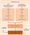
Figure 1.
Figure 1.. Identification of germline CDKN2A variant of uncertain significance (VUS) for functional characterization.
Workflow to identify the 29 germline CDKN2A VUS selected for characterization in our functional assay. CDKN2A VUSs were classified using American College of Medical Genetics (ACMG) guidelines and either identified in patients with pancreatic ductal adenocarcinoma (PDAC) from published literature or in ClinVar.
Figure 2.
Figure 2.. Development and validation of functional assay.
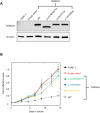
(A) Western blot of whole cell lysates from PANC-1 cells and PANC-1 cells stably expressing wild type CDKN2A (p16INK4A) or selected pathogenic variants using anti-CDKN2A and anti-vinculin antibodies as a loading control. No expression of CDKN2A (p16INK4A) detected in PANC-1 cells or PANC-1 cell stably expressing empty vector. CDKN2A (p16INK4A) expression detected in PANC-1 cells stably expressing pathogenic variants. (B) Growth of PANC-1 cell and PANC-1 cells stably expressing wild type CDKN2A cDNA (p16INK4A) or selected pathogenic variants over 14 days in culture. Cell proliferation values are shown as mean (n = 3) ± s.d. normalized to PANC-1 cell stably expressed with empty vector. Significant growth inhibition in PANC-1 cell stably expressing wild type CDKN2A (p16INK4A). *p < 0.01. Original files of the full raw unedited blots and the uncropped blots with the relevant CDKN2A and vinculin bands presented in Figure 2—source data 1, Figure 2—source data 2 and 3. For raw data in Figure 2, please refer to Figure 2—source data 4.
Figure 3.
Figure 3.. Functional characterization of CDKN2A variants.
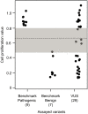
Functional characterization of 45 CDKN2A variants (9 pathogenic variants, 7 benign variants, 29 variants of uncertain significance [VUSs]) in PANC-1 cells. Cell proliferation values are shown as mean (n = 3) ± s.d. normalized to PANC-1 cell stably expressed with empty vector. Assayed variants with mean cell proliferation values: (1) above the gray shaded area (>0.81) were classified as functionally deleterious, (2) below the gray shaded area (<0.44) were classified as functionally benign, (3) in the gray area above the threshold dotted line (≥0.66 but ≤0.81) were potentially functionally deleterious, and (4) in the gray area below the threshold dotted line (<0.66 but ≥0.44) were potentially functionally neutral. For raw data in this figure, please refer to Figure 3—source data 1.
Figure 3—figure supplement 1.
Figure 3—figure supplement 1.. CDKN2A expression in human pancreatic ductal adenocarcinoma (PDAC) cells.
Western blot of whole cell lysates from 293T cells, a human embryonic kidney cell line and PANC-1, MIA PaCa-2, and AsPC-1 cells, human PDAC cell lines using anti-CDKN2A and anti-vinculin antibodies as a loading control. No expression of CDKN2A (p16INK4A) detected in human PDAC cells. CDKN2A (p16INK4A) expression detected in 293T cells. For the original files of the full raw unedited blots and figures with the uncropped blots with the relevant bands clearly labelled, please refer to Figure 3—figure supplement 1—source data 1, Figure 3—figure supplement 1—source data 2, Figure 3—figure supplement 1—source data 3.
Figure 3—figure supplement 2.
Figure 3—figure supplement 2.. Functional characterization of CDKN2A variants in MIA PaCa-2 and AsPC-1 cells.
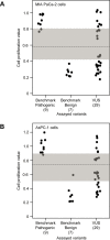
Functional characterization of 45 CDKN2A variants (9 pathogenic variants, 7 benign variants, 29 variants of uncertain significance [VUSs]). Cell proliferation values are shown as mean (n = 3) ± s.d. normalized to MIA PaCa-2 (A) or AsPc-1 (B) cell stably expressed with empty vector. Assayed variants with mean cell proliferation values: (1) above the gray shaded area (>0.80 for MIA PaCa-2 cells and 0.91 for AsPC-1 cells) were classified as functionally deleterious, (2) below the gray shaded area (<0.45 for MIA PaCa-2 cells and 0.51 for AsPC-1 cells) were classified as functionally benign, (3) in the gray area above the threshold dotted line (≥0.57 but ≤0.80 for MIA PaCa-2 cells and ≥0.74 but ≤0.91 for AsPC-1 cells) were potentially functionally deleterious, and (4) in the gray area below the threshold dotted line (<0.57 but ≥0.45 for MIA PaCa-2 cells and <0.74 but ≥0.51 for AsPC-1 cells) were potentially functionally neutral. For raw data in this figure, please refer to Figure 3—figure supplement 2—source data 1.
Figure 3—figure supplement 3.
Figure 3—figure supplement 3.. Functional characterization of CDKN2A variants.
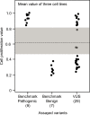
Functional characterization of 45 CDKN2A variants (9 pathogenic variants, 7 benign variants, 29 variants of uncertain significance [VUSs]). Cell proliferation values are shown as mean of PANC-1, MIA PaCa-2, and AsPc-1 cell values. Assayed variants with mean cell proliferation values: (1) above the gray shaded area (>0.83) were classified as functionally deleterious, (2) below the gray shaded area (<0.47) were classified as functionally benign, (3) in the gray area above the threshold dotted line (≥0.62 but ≤0.83) were potentially functionally deleterious, and (4) in the gray area below the threshold dotted line (<0.62 but ≥0.47) were potentially functionally neutral. For raw data in this figure, please refer to Figure 3—figure supplement 3—source data 1.
Figure 4.
Figure 4.. Cell cycle analysis of CDKN2A variants.
G1 percent for 45 CDKN2A variants (9 pathogenic variants, 7 benign variants, 29 variants of uncertain significance [VUSs]) in PANC-1 cells. Significant inhibition of G1 arrest function of CDKN2A by benchmark pathogenic variants and functionally deleterious or potentially functionally deleterious VUSs. *p < 0.01. For raw data in this figure, please refer to Figure 4 and Table 1.
Figure 4—figure supplement 1.
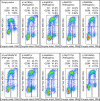
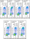
Figure 4—figure supplement 1.. Cell cycle analysis of benchmark pathogenic CDKN2A variants.
Cell cycle analysis was performed with percentages of cells in each phase (G1, G2_M, and S) indicated. Flow cytometry analysis images for PANC-1 cell stably expressed with empty vector or benchmark pathogenic variants.
Figure 4—figure supplement 2.
Figure 4—figure supplement 2.. Cell cycle analysis of benchmark benign CDKN2A variants.
Cell cycle analysis was performed with percentages of cells in each phase (G1, G2_M, and S) indicated. Flow cytometry analysis images for PANC-1 cell stably expressed with empty vector or benchmark benign variants.
Figure 4—figure supplement 3.
Figure 4—figure supplement 3.. Cell cycle analysis of functionally deleterious and potentially functionally deleterious CDKN2A variants of uncertain significance (VUSs).
Cell cycle analysis was performed with percentages of cells in each phase (G1, G2_M, and S) indicated. Flow cytometry analysis images for PANC-1 cell stably expressed with empty vector or functionally deleterious and potentially functionally deleterious VUSs.
Figure 4—figure supplement 4.
Figure 4—figure supplement 4.. Cell cycle analysis of functionally neutral and potentially functionally neutral CDKN2A variants of uncertain significance (VUSs).
Cell cycle analysis was performed with percentages of cells in each phase (G1, G2_M, and S) indicated. Flow cytometry analysis images for PANC-1 cell stably expressed with empty vector or functionally neutral and potentially functionally neutral VUSs.
Figure 4—figure supplement 5.
Figure 4—figure supplement 5.. G2_M percent of CDKN2A variants expression cells.
G2_M percent for 45 CDKN2A variants (9 pathogenic variants, 7 benign variants, 29 variants of uncertain significance [VUS]) in PANC-1 cells. G2_M percent in benchmark pathogenic variants and functionally deleterious or potentially functionally deleterious VUSs expression cells were significantly higher than that in benchmark benign variants expression cells. *p < 0.01. For raw data in this figure, please refer to Figure 4 and 1.

Similar articles

Cited by

References

    1. Bouvet D, Bodo S, Munier A, Guillerm E, Bertrand R, Colas C, Duval A, Coulet F, Muleris M. Methylation Tolerance-Based Functional Assay to Assess Variants of Unknown Significance in the MLH1 and MSH2 Genes and Identify Patients With Lynch Syndrome. Gastroenterology. 2019;157:421–431. doi: 10.1053/j.gastro.2019.03.071. - DOI - PubMed
    1. Brand R, Borazanci E, Speare V, Dudley B, Karloski E, Peters MLB, Stobie L, Bahary N, Zeh H, Zureikat A, Hogg M, Lee K, Tsung A, Rhee J, Ohr J, Sun W, Lee J, Moser AJ, DeLeonardis K, Krejdovsky J, Dalton E, LaDuca H, Dolinsky J, Colvin A, Lim C, Black MH, Tung N. Prospective study of germline genetic testing in incident cases of pancreatic adenocarcinoma. Cancer. 2018;124:3520–3527. doi: 10.1002/cncr.31628. - DOI - PubMed
    1. Caldas C, Hahn SA, da Costa LT, Redston MS, Schutte M, Seymour AB, Weinstein CL, Hruban RH, Yeo CJ, Kern SE. Frequent somatic mutations and homozygous deletions of the p16 (MTS1) gene in pancreatic adenocarcinoma. Nature Genetics. 1994;8:27–32. doi: 10.1038/ng0994-27. - DOI - PubMed
    1. Canto MI, Harinck F, Hruban RH, Offerhaus GJ, Poley JW, Kamel I, Nio Y, Schulick RS, Bassi C, Kluijt I, Levy MJ, Chak A, Fockens P, Goggins M, Bruno M, International Cancer of Pancreas Screening CAPS Consortium International Cancer of the Pancreas Screening (CAPS) Consortium summit on the management of patients with increased risk for familial pancreatic cancer. Gut. 2013;62:339–347. doi: 10.1136/gutjnl-2012-303108. - DOI - PMC - PubMed
    1. Canto M.I, Almario JA, Schulick RD, Yeo CJ, Klein A, Blackford A, Shin EJ, Sanyal A, Yenokyan G, Lennon AM, Kamel IR, Fishman EK, Wolfgang C, Weiss M, Hruban RH, Goggins M. Risk of Neoplastic Progression in Individuals at High Risk for Pancreatic Cancer Undergoing Long-term Surveillance. Gastroenterology. 2018;155:740–751. doi: 10.1053/j.gastro.2018.05.035. - DOI - PMC - PubMed

Publication types

MeSH terms

Substances