. 2021 Dec 7:27:666-678.
eCollection 2021.

Collagen matrix perturbations in corneal stroma of Ossabaw mini pigs with type 2 diabetes

Affiliations

Collagen matrix perturbations in corneal stroma of Ossabaw mini pigs with type 2 diabetes

Nishant R Sinha et al. Mol Vis. .

Abstract

Purpose: Diabetes mellitus (DM) is a metabolic disorder that affects over 450 million people worldwide. DM is characterized by hyperglycemia, causing severe systemic damage to the heart, kidneys, skin, vasculature, nerves, and eye. Type 2 diabetes (T2DM) constitutes 90% of clinical cases and is the most common cause of blindness in working adults. Also, about 70% of T2DM patients show corneal complications including delayed wound healing, often described as diabetic keratopathy (DK). Despite the increasing severity of DM, the research on DK is bleak. This study investigated cellular morphology and collagen matrix alterations of the diabetic and non-diabetic corneas collected from Ossabaw mini pigs, a T2DM animal model with a "thrifty genotype."

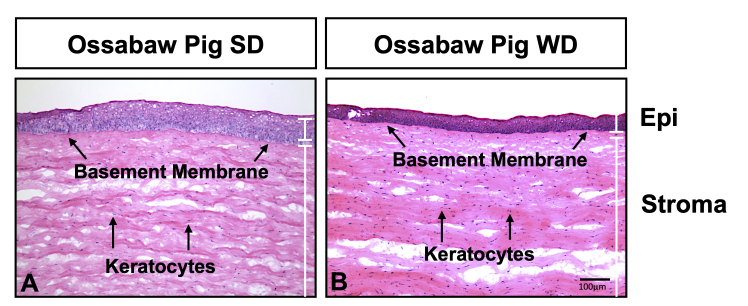
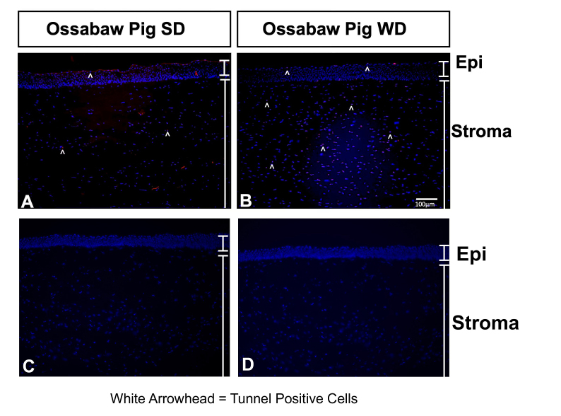
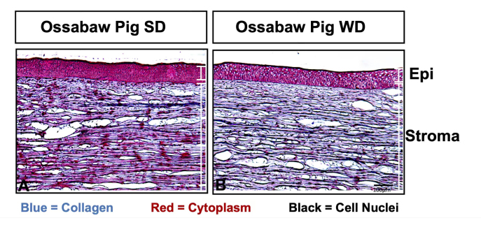
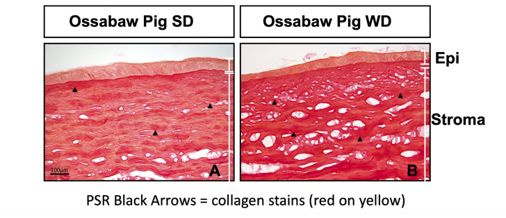
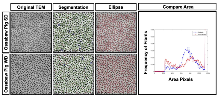
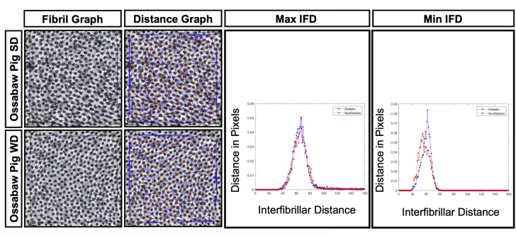
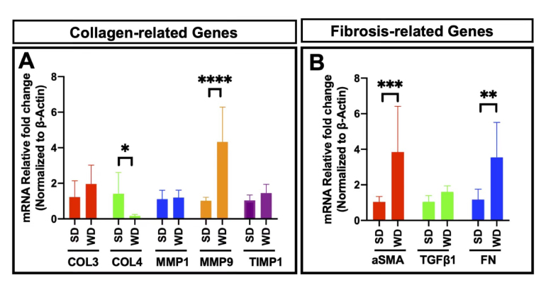
Methods: Pig corneas were collected from six-month-old Ossabaw miniature pigs fed on a western diet (WD) for ten weeks. The tissues were processed for immunohistochemistry and analyzed using hematoxylin and eosin staining, Mason Trichrome staining, Picrosirus Red staining, Collage I staining, and TUNEL assay. mRNA was prepared to quantify fibrotic gene expression using quantitative reverse-transcriptase PCR (qRT-PCR). Transmission electron microscopy (TEM) was performed to evaluate stromal fibril arrangements to compare collagen dynamics in WD vs. standard diet (SD) fed Ossabaw pig corneas.

Results: Ossabaw mini pigs fed on a WD for 10 weeks exhibit classic symptoms of metabolic syndrome and hyperglycemia seen in T2DM patients. We observed significant disarray in cornea stromal collagen matrix in Ossabaw mini pigs fed on WD compared to the age-matched mini pigs fed on a standard chow diet using Masson Trichome and Picrosirius Red staining. Furthermore, ultrastructure evaluation using TEM showed alterations in stromal collagen fibril size and organization in diabetic corneas compared to healthy age-matched corneas. These changes were accompanied by significantly decreased levels of Collagen IV and increased expression of matrix metallopeptidase 9 in WD-fed pigs.

Conclusions: This pilot study indicates that Ossabaw mini pigs fed on WD showed collagen disarray and altered gene expression involved in wound healing, suggesting that corneal stromal collagens are vulnerable to diabetic conditions.


Figures

Figure 1
Figure 1
Hematoxylin and Eosin staining suggest an increase in keratocytes (black arrows) in the Western diet (WD) fed Ossabaw mini pigs (B) compared to the Standard diet (SD) fed pigs (A). Green arrows indicate the basement membrane.
Figure 2
Figure 2
Tunnel assay indicates an increase in Tunnel-positive cells (white arrowheads) in the Western diet (WD) fed Ossabaw mini pigs (B) compared to the Standard diet (SD) fed pigs (A). Panels C and D are negative controls where Tdt enzyme and Anti-Digoxigenin Conjugate were respectively excluded during the staining procedure. Arrowheads indicate the apoptotic cells.
Figure 3
Figure 3
Mason trichome staining depicts cell nuclei (black), cytoplasm (red), and collagens (blue). The Western diet (WD) fed Ossabaw mini pigs (B) show decreased cytoplasm staining and a less even blue stain than the Standard diet (SD) fed pigs (A).
Figure 4
Figure 4
Immunohistochemistry of Collagen I (Col I) displays increased collagen aggregates (yellow triangles) and an increased Col I expression at the basement membrane (white arrows) in the Western diet (WD) fed Ossabaw mini pigs (B) compared to the Standard diet (SD) fed pigs (A). Panels C and D are negative controls where primary and secondary antibodies were excluded during antibody staining procedure.
Figure 5
Figure 5
Picrosirius red (PSR) stains collagen in the corneal stroma. A red-on-yellow stain signifies collagen type 1. PSR stained Western diet (WD) fed pig corneal section (B) shows high numbers of irregular collagen compared to the Standard diet (SD) fed pigs (A).
Figure 6
Figure 6
Quantitation of transmission electron microscopy of collagen fibrils in the western diet (WD) fed and Standard diet (SD) fed Ossabaw mini pigs. Panels A and D are the original transverse section of the transmission electron microscopy images taken at 50,000 magnification. Panels B and F are segmentations that identify all the collagens present in the figure. Panels C and G are individual red ellipses made around collagen fibrils used to quantify the size of fibrils. Panel H is a graphical representation of collagen fibril sizes found in the image. Western diet pigs show an overall collagen size difference compared to standard diet pigs.
Figure 7
Figure 7
Quantitation of Transmission Electron Microscopy of collagen fibrils in the Western diet (WD) fed and Standard diet (SD) fed Ossabaw mini pigs. A and C are fibril graphs that show each connection between collagen fibrils. B and D show the minimum distance between collagen fibrils (red) and the maximum distance between fibrils (blue). E is a graphical representation of maximum interfibrillar distance and F is a graphical representation of minimum interfibrillar distance.
Figure 8
Figure 8
Gene expression in the Western diet (WD) fed and Standard diet (SD) fed Ossabaw pig corneas. A represents collagen-related genes- collagen 3 (COL 3), collagen 4 (COL 4), metalloproteases 1 (MMP1), metalloproteases 9 (MMP9), and tissue inhibitor of metalloprotease 1 (TIMP1). B represents fibrosis-related genes alpha-smooth muscle action (ɑSMA), transformation growth factor beta 1 (TGFβ1), and fibronectin (FN). The WD-fed Ossabaw mini pigs corneas had significant alterations in COL4, MMP9, ɑSMA, TGFβ1, and FN. compared to the SD-fed pigs (P, *<0.05, **<0.01, ***<0.001, ****<0.0001).

Similar articles

Cited by

References

    1. Lee SY, Mesfin FB. Blindness. In: StatPearls. Elsevier Inc.; 2020. doi:10.1016/B978-0-12-803678-5.00036-910.1016/B978-0-12-803678-5.00036-9 - DOI - DOI
    1. Schmidt AM. Highlighting Diabetes Mellitus. Arterioscler Thromb Vasc Biol. 2018;38:e1–8. - PMC - PubMed
    1. Copeland RA, Natalie A. Copeland and Afshari’s Principles and Practice of Cornea. 1st edition. Jaypee Brothers Medical Publishers (P) Ltd; 2013. 1460 p.
    1. Bullard KM, Cowie CC, Lessem SE, Saydah SH, Menke A, Geiss LS, Orchard TJ, Rolka DB, Imperatore G. Prevalence of Diagnosed Diabetes in Adults by Diabetes Type — United States, 2016. MMWR Morb Mortal Wkly Rep. 2018;67:359–61. - PMC - PubMed
    1. Negi A, Vernon SA. An overview of the eye in diabetes. In: Journal of the Royal Society of Medicine. Vol 96. Royal Society of Medicine Press Ltd; 2003:266–272. doi:10.1258/jrsm.96.6.26610.1258/jrsm.96.6.266 - DOI - DOI - PMC - PubMed

Publication types

Substances