

Qualitative Alterations on Corneal Endothelial Cell Morphometry and Hexagonality After Cataract Surgery
- PMID: 35002220
- PMCID: PMC8721951
- DOI: 10.2147/OPTH.S338001
Qualitative Alterations on Corneal Endothelial Cell Morphometry and Hexagonality After Cataract Surgery
Abstract
Aim: To deeply analyze quantitative and qualitative changes of corneal endothelium after longitudinal phacoemulsification.
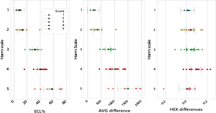
Methods: In this prospective interventional case series study, 50 eyes with age-related cataract have been evaluated preoperatively, intraoperatively and postoperatively. The measured parameters were surgically induced endothelial cell loss (ECL), average endothelial cell area (AVG) and hexagonality (HEX).
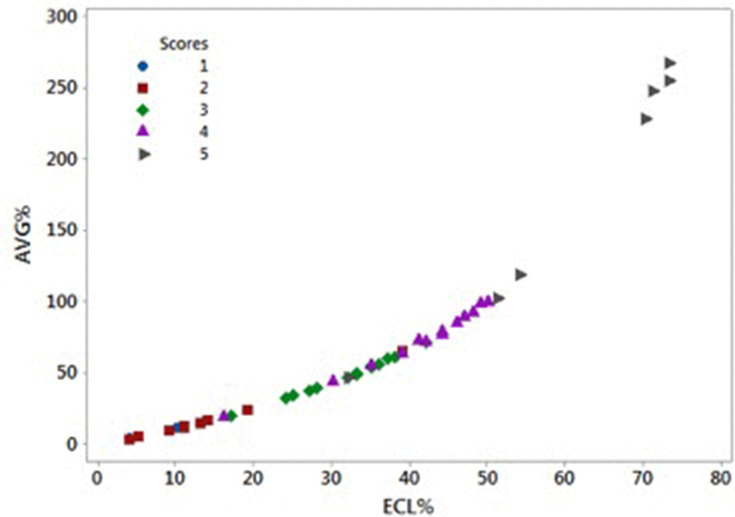
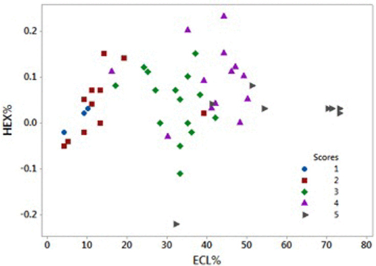
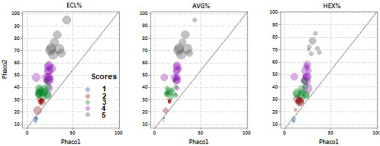
Results: The relationship among the measured parameters and the energy dissipated during the two phases of phacoemulsification, sculpting and quadrant removal, has been analyzed with regard to the 5-score harm scale, a new method suggested to categorize cataracts. Two phases of phacoemulsification are linearly related (r = 1.5, P < 0.001, r2 = 79%). Plus, a quadratic model described the correlation between the percentages of ECL and AVG (P < 0.0001), while there was no specific model for the correlation between ECL and HEX.
Conclusion: The 5-score harm scale allows to predict the mean changes in percentages of ECL, AVG and HEX of endothelial cells after longitudinal phacoemulsification. Also, this study confirms that the main damage on corneal endothelium is due to the energy delivered in the second phase of phacoemulsification.
Keywords: average cell area; cataract; endothelial cell loss; hexagonality; phacoemulsification.
© 2021 Sorrentino.
Conflict of interest statement
The author reports no conflicts of interest.
Figures
Similar articles
-
A Pilot Study to Propose a "Harm Scale", a New Method to Predict Risk of Harm to the Corneal Endothelium Caused by Longitudinal Phacoemulsification, and the Subsequent Effect of Endothelial Damage on Post Operative Visual Acuity.PLoS One. 2016 Jan 13;11(1):e0146580. doi: 10.1371/journal.pone.0146580. eCollection 2016. PLoS One. 2016. PMID: 26761198 Free PMC article.
-
Torsional phacoemulsification: A pilot study to revise the "harm scale" evaluating the endothelial damage and the visual acuity after cataract surgery.PLoS One. 2017 Oct 26;12(10):e0186975. doi: 10.1371/journal.pone.0186975. eCollection 2017. PLoS One. 2017. PMID: 29073200 Free PMC article. Clinical Trial.
-
Corneal endothelial cell loss after cataract extraction by using ultrasound phacoemulsification versus a fluid-based system.Cornea. 2008 Jan;27(1):17-21. doi: 10.1097/ICO.0b013e3181583115. Cornea. 2008. PMID: 18245961 Clinical Trial.
-
Endothelial cell loss and refractive predictability in femtosecond laser-assisted cataract surgery compared with conventional cataract surgery.Acta Ophthalmol. 2014 Nov;92(7):617-22. doi: 10.1111/aos.12406. Epub 2014 Jun 2. Acta Ophthalmol. 2014. PMID: 24888390 Clinical Trial.
-
Advances in cataract surgery: preserving the corneal endothelium.Curr Opin Ophthalmol. 2015 Jan;26(1):22-7. doi: 10.1097/ICU.0000000000000121. Curr Opin Ophthalmol. 2015. PMID: 25415300 Review.
Cited by
-
Effect of anterior chamber depth on corneal endothelium following phacoemulsification among patients with different axial lengths.Int Ophthalmol. 2025 Jan 29;45(1):45. doi: 10.1007/s10792-025-03415-7. Int Ophthalmol. 2025. PMID: 39881004
-
Corneal Endothelial Characteristics in Normal Chinese Han Children and Youngsters: A Study from the Specular Microscopy Descriptions.Dis Markers. 2022 May 20;2022:5338725. doi: 10.1155/2022/5338725. eCollection 2022. Dis Markers. 2022. PMID: 35634448 Free PMC article.
References
-
- Bourne RR, Minassian DC, Dart JK, Rosen P, Kaushal S, Wingate N. Effect of cataract surgery on the corneal endothelium: modern phacoemulsification compared with extracapsular cataract surgery. Ophthalmology. 2004;111:679–685. - PubMed
LinkOut - more resources
Full Text Sources
