. 2021 Dec 10:1076:151-182.
doi: 10.3897/zookeys.1076.69025. eCollection 2021.

Gems of the southern Japanese seas - four new species of Edwardsianthus (Anthozoa, Actiniaria, Edwardsiidae) with redescriptions of two species

Affiliations

Gems of the southern Japanese seas - four new species of Edwardsianthus (Anthozoa, Actiniaria, Edwardsiidae) with redescriptions of two species

Takato Izumi et al. Zookeys. .

Abstract

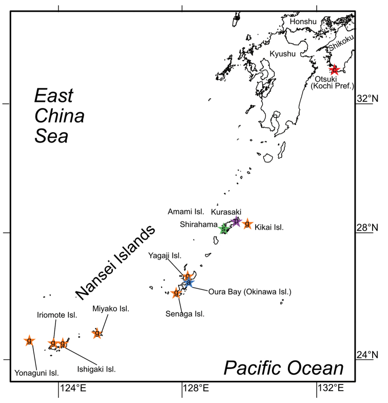
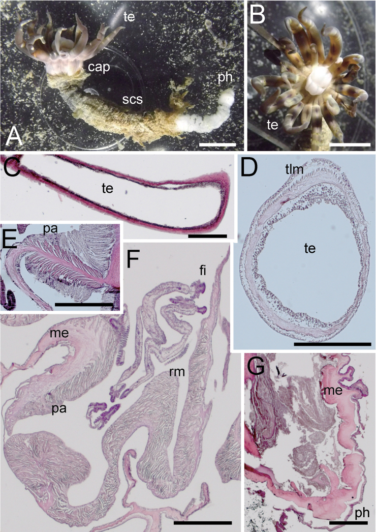
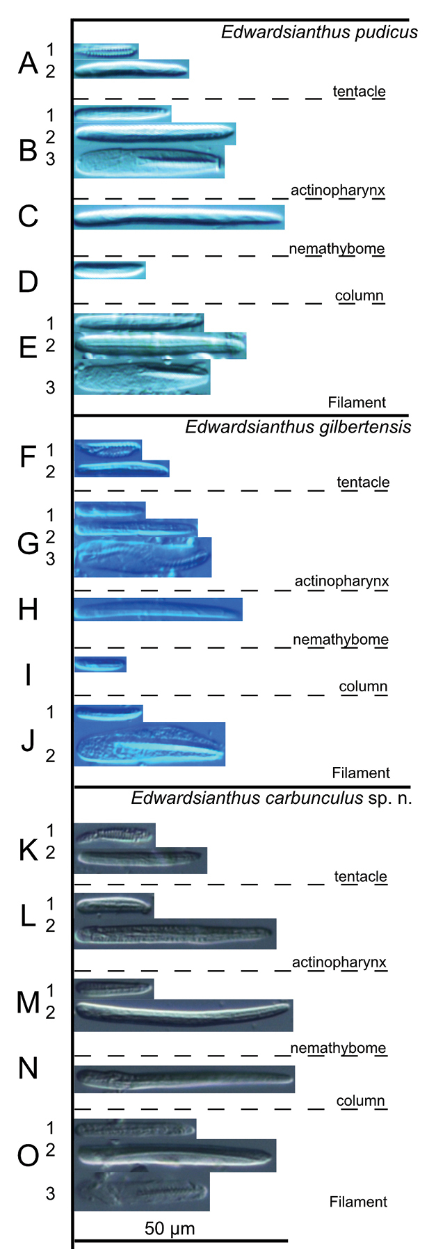
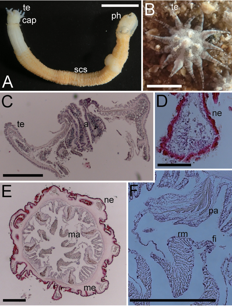
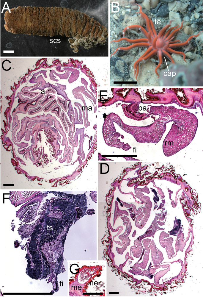
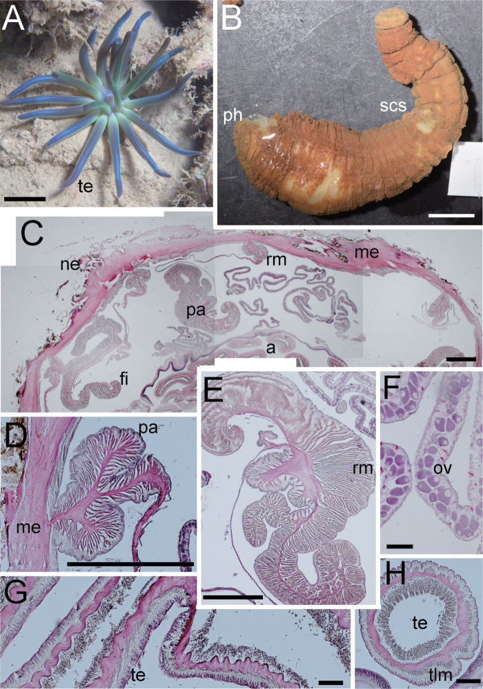
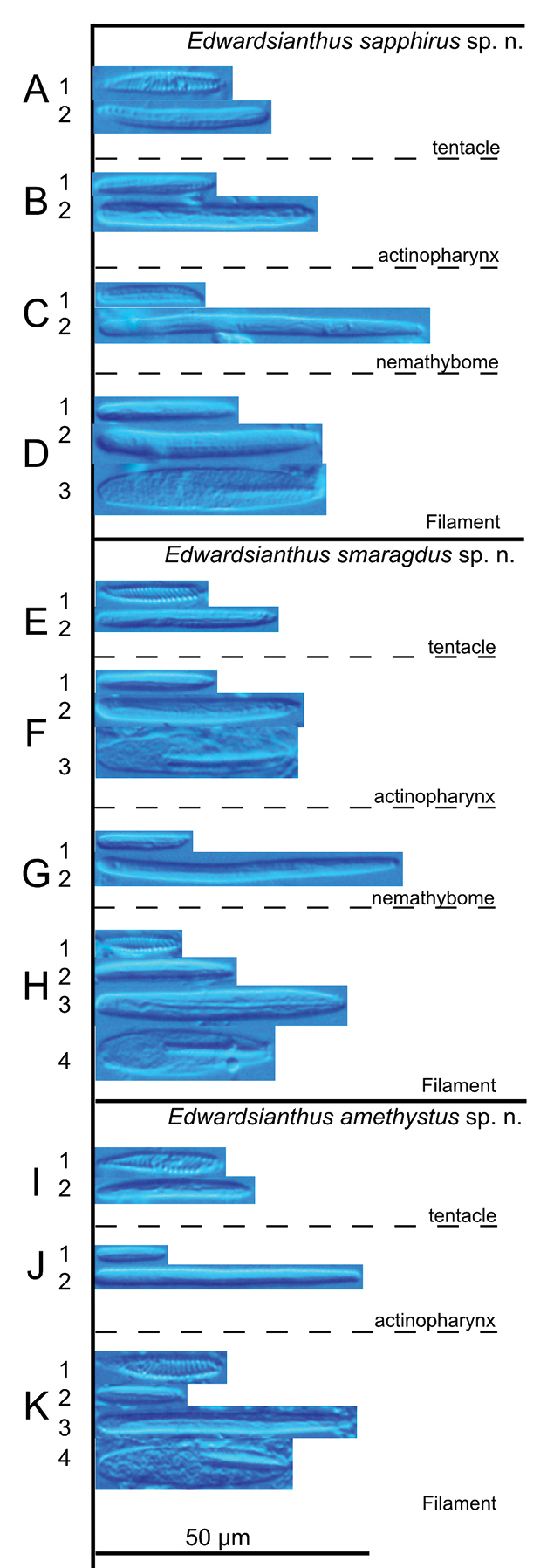
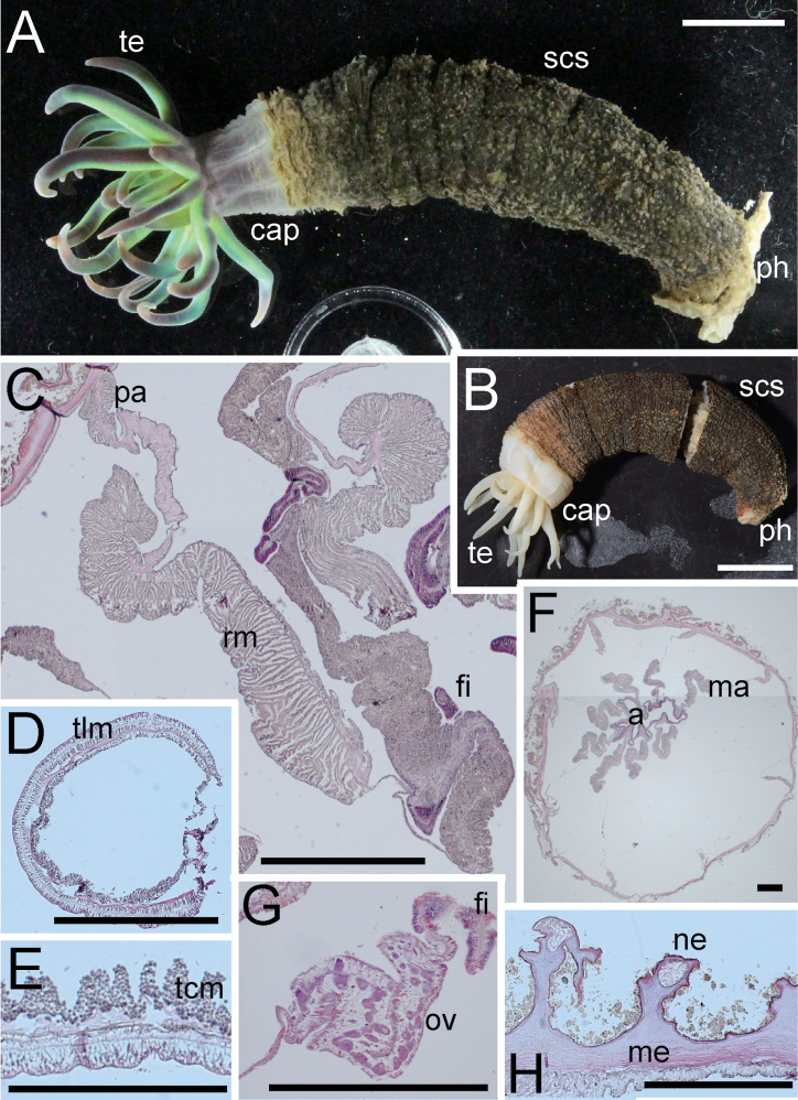
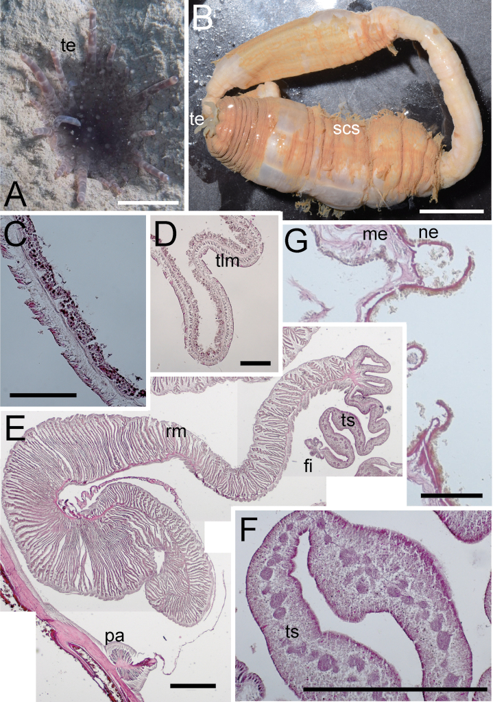
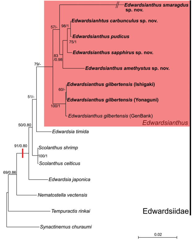
Edwardsianthus England, 1987 is a genus of Edwardsiidae, a family of burrowing and worm-like sea anemones characterized by lacking four mesenteries in the first cycle and containing only one type of nematocysts in nemathybomes. Until now, this genus has accommodated only two species since its establishment and has been recorded only from Indo-West Pacific regions. In this study, six species are reported from Japan: two are previously known species, E.pudicus (Klunzinger, 1877) and E.gilbertensis (Carlgren, 1931); four are new species, E.carbunculus sp. nov., E.sapphirus sp. nov., E.smaragdus sp. nov., and E.amethystus sp. nov. Based on these results, the diagnostic features of the genus are revised.

Keywords: Cnidaria; Kochi; Nansei Islands; Pacific Ocean; cnidae; nemathybomes; northernmost distribution limit; phylogeny; taxonomy.


Figures

Figure 1.
Figure 1.
Localities of the specimens of the Edwardsianthus species collected in this study. Purple star marked by p indicates collection locality of Edwardsianthuspudicus; orange stars are those of E.gilbertensis; red star marked by c is E.carbunculus sp. nov.; blue star marked by a is E.sapphirus sp. nov.; green star marked by s is E.smaragdus sp. nov.
Figure 2.
Figure 2.
External and internal morphology of Edwardsianthuspudicus (NSMT-Co 1702) A outer view of living specimen B oral view of living specimen C longitudinal section of tentacle D transverse section of tentacle E transverse section of parietal muscle F transverse section of macrocnemes G longitudinal section of physa. Abbreviations: cap, capitulum; fi, filament; me, mesoglea; pa, parietal muscle; ph, physa; rm, retractor muscle; scs, scapus; te, tentacle; tlm, tentacular longitudinal muscle. Scale bars: 5 mm (A, B); 1 mm (C); 500 µm (D, F, G); 100 µm (E).
Figure 3.
Figure 3.
Cnidae of Edwardsianthus species A–EE.pudicus (NSMT-Co 1702) A1 spirocyst in tentacle; A2 basitrich in tentacle B1 small basitrich in actinopharynx B2 large basitrich in actinopharynx B3 microbasic p-mastigophore in actinopharynx C basitrich in nemathybome D basitrich in column E1 small basitrich in filament E2 large basitrich in filament E3 microbasic p- mastigophore in filament F–JE.gilbertensis (CMNH-ZG 06527) F1 spirocyst in tentacle F2 basitrich in tentacle G1 small basitrich in actinopharynx G2 large basitrich in actinopharynx G3 microbasic p-mastigophore in actinopharynx H basitrich in nemathybome I basitrich in nemathybome J1 basitrich in filament J2 microbasic p- mastigophore in filament K–OE.carbunculus sp. nov. (CMNH-ZG 05954) K1 spirocyst in tentacle K2 basitrich in tentacle L1 small basitrich in actinopharynx L2 large basitrich in actinopharynx M1 small basitrich in nemathybome M2 large basitrich in nemathybome N basitrich in column O1 small basitrich in filament O2 large basitrich in filament O3 microbasic p-mastigophore in filament.
Figure 4.
Figure 4.
External and internal morphology of Edwardsianthusgilbertensis (CMNH-ZG 06527 except for B) A outer view of preserved specimen B oral view of living specimen with 18 tentacles in the habitat C longitudinal section of oral end D transverse section of nemathybome E transverse section of column in lower part; F. Enlarged view of transverse section of mesenteries. Abbreviations: a, actinopharynx; cap, capitulum; fi, filament; ma, macrocneme; ne, nemathybomes; pa, parietal muscle; ph, physa; rm, retractor muscle; scs, scapus; te, tentacle. Scale bars: 5 mm (A); 1 mm (B); 500 µm (C, E, F); 100 µm (D). Photograph B by Kensuke Yanagi.
Figure 5.
Figure 5.
External and internal morphology of Edwardsianthuscarbunculus sp. nov. (CMNH-ZG 05954) A outer view of preserved specimen (aboral end is damaged) B oral view of living specimen in the habitat (photograph Kensuke Yanagi) C transverse section of column in upper part D transverse section of lower column in lower part E transverse section of the macrocneme F transverse section of testis G transverse section of nemathybomes. Abbreviations: a, actinopharynx; cap, capitulum; fi, filament; me, mesoglea; ne, nemathybomes; pa, parietal muscle; rm, retractor muscle; scs, scapus; te, tentacle. Scale bars: 5 mm (A, B); 500 µm (C–F); 100 µm (G). Picture B taken by Kensuke Yanagi.
Figure 6.
Figure 6.
External and internal morphology of Edwardsianthussapphirus sp. nov. (CMNH-ZG 09761). A oral view of living specimen in the habitat B outer view of preserved specimen; C. Transverse section of column in upper part D enlarged view of transverse section of parietal muscle E enlarged view of transverse section of retractor muscle F transverse section of ovary G longitudinal section of tentacle H transverse section of tentacle. Abbreviations: a, actinopharynx; fi, filament; me, mesoglea; ne, nemathybome; oo, oocytes; pa, parietal muscle; ph, physa; rm, retractor muscle; scs, scapus; te, tentacle; tlm, tentacular longitudinal muscle. Scale bars: 1 cm (A, B); 500 µm (C–E); 100 µm (F–H).
Figure 7.
Figure 7.
Cnidae of Edwardsianthus species A–DE.sapphirus sp. nov. (CMNH-ZG 09761) A1 spirocyst in tentacle A2 basitrich in tentacle B1 small basitrich in actinopharynx B2 large basitrich in actinopharynx C1 small basitrich in nemathybome C2 large basitrich in nemathybome D1 small basitrich in filament D2 large basitrich in filament D3 microbasic p-mastigophore in filament E–HE.smaragdus sp. nov. (CMNH-ZG 09762) E1 spirocyst in tentacle E2 basitrich in tentacle F1 small basitrich in actinopharynx F2 large basitrich in actinopharynx F3 microbasic p-mastigophore in actinopharynx G1 small basitrich in nemathybome; G2 large basitrich in nemathybome; H1 spirocyst in filament; H2 small basitrich in filament H3 large basitrich in filament H4 microbasic p-mastigophore in filament I–KE.amethystus sp. nov. (CMNH-ZG 09763) I1 spirocyst in tentacle I2 basitrich in tentacle J1 small basitrich in actinopharynx J2 large basitrich in actinopharynx K1 spirocyst in filament K2 small basitrich in filament K3 large basitrich in filament K4 microbasic p-mastigophore in filament.
Figure 8.
Figure 8.
External and internal morphology of Edwardsianthussmaragdus sp. nov. (CMNH-ZG 09762). A outer view of living specimen B outer view of preserved specimen C transverse section of retractor muscle D transverse section of the tentacle E longitudinal section of the tentacle F transverse section of column G enlarged view of transverse section of ovary H transverse section of a nemathybome. Abbreviations: a, actinopharynx; cap, capitulum; fi, filament; ma, macrocneme; me, mesoglea; ne, nemathybomes; ov, ovary; pa, parietal muscle; ph, physa; rm, retractor muscle; scs, scapus; te, tentacle; tcm, tentacular circular muscle; tlm, tentacular longitudinal muscle. Scale bars: 1 cm (A, B); 500 µm in (CH).
Figure 9.
Figure 9.
External and internal morphology of Edwardsianthusamethystus sp. nov. (CMNH-ZG 09763) A oral view of living specimen in the habitat B outer view of preserved specimen C longitudinal section of tentacle D transverse section of tentacle E transverse section of retractor muscle F transverse section of testis G transverse section of trace of nemathybome. Abbreviations: fi, filament; me, mesoglea; ne, nemathybome-like structure; pa, parietal muscle; rm, retractor muscle; scs, scapus; te, tentacle; tlm, tentacle longitudinal muscle; ts, testis. Scale bars: 1 cm (A, B); 500 µm (E, F); 100 µm (C, D, G).
Figure 10.
Figure 10.
Maximum-likelihood tree of the order Actiniaria based on the combined dataset of mitochondrial 12S and 16S and nuclear 18S rDNA (total 2866 bp). The clade of the genus Edwardsianthus is colored in a red box. Red bar at node indicates the position at which nemathybomes would be obtained. Numbers indicate ML bootstrap support values followed by BI posterior probabilities of the nodes (bootstrap values of ≥ 50% and posterior probabilities ≥ 0.5 are shown).

Similar articles

References

    1. Andres A. (1881) Prodromus neapolitanae actiniarum faunae addito generalis actiniarum bibliographiae catalogo. Mitteilungen aus der Zoologischen Station zu Neape 2: 305–371.
    1. Apakupakul K, Siddall ME, Burreson EM. (1999) Higher level relationships of leeches (Annelida: Clitellata: Euhirudinea) based on morphology and gene sequences. Molecular Phylogenetics and Evolution 12: 350–359. 10.1006/mpev.1999.0639 - DOI - PubMed
    1. Carlgren O. (1892) Beiträge zur Kenntnis der Edwardsien. Öfversigt af Kongliga Vetenskaps-Akademiens Förhandlingar 1892: 451–461.
    1. Carlgren O. (1921) Actiniaria Part I. Danish Ingolf-Expedition 5: 1–241.
    1. Carlgren O. (1931) Zur Kenntnis der Actiniaria Abasilaria. Arkiv für Zoologi 23: 1–48.

LinkOut - more resources