

Novel biosynthesis, characterization and bio-catalytic potential of green algae (Spirogyra hyalina) mediated silver nanomaterials
- PMID: 35002436
- PMCID: PMC8717159
- DOI: 10.1016/j.sjbs.2021.09.013
Novel biosynthesis, characterization and bio-catalytic potential of green algae (Spirogyra hyalina) mediated silver nanomaterials
Abstract
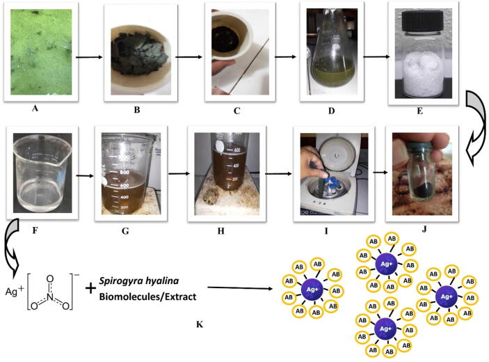
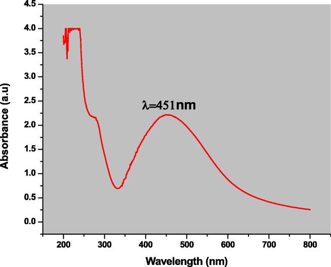
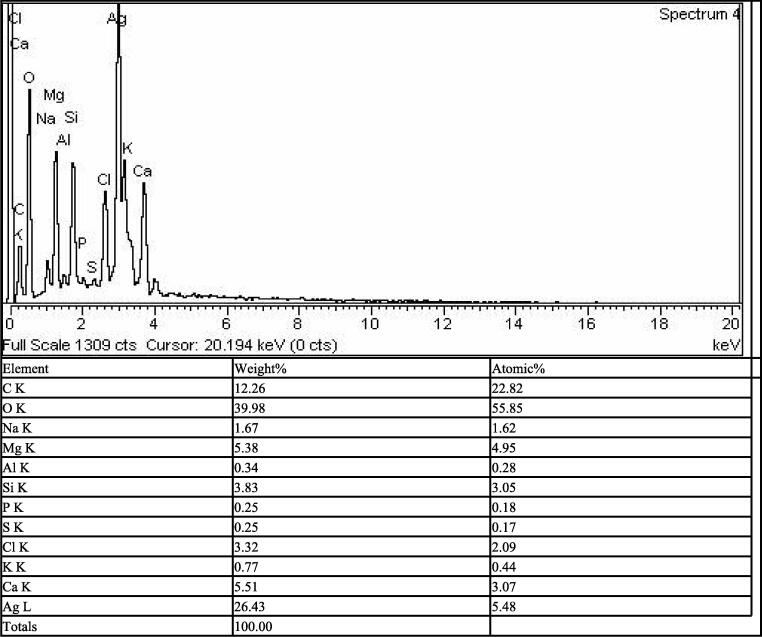
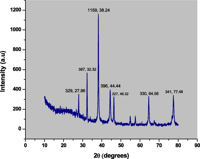
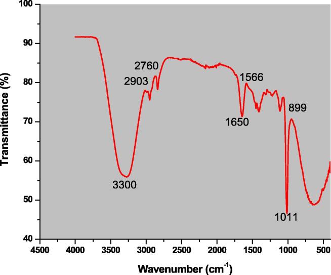
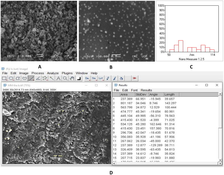
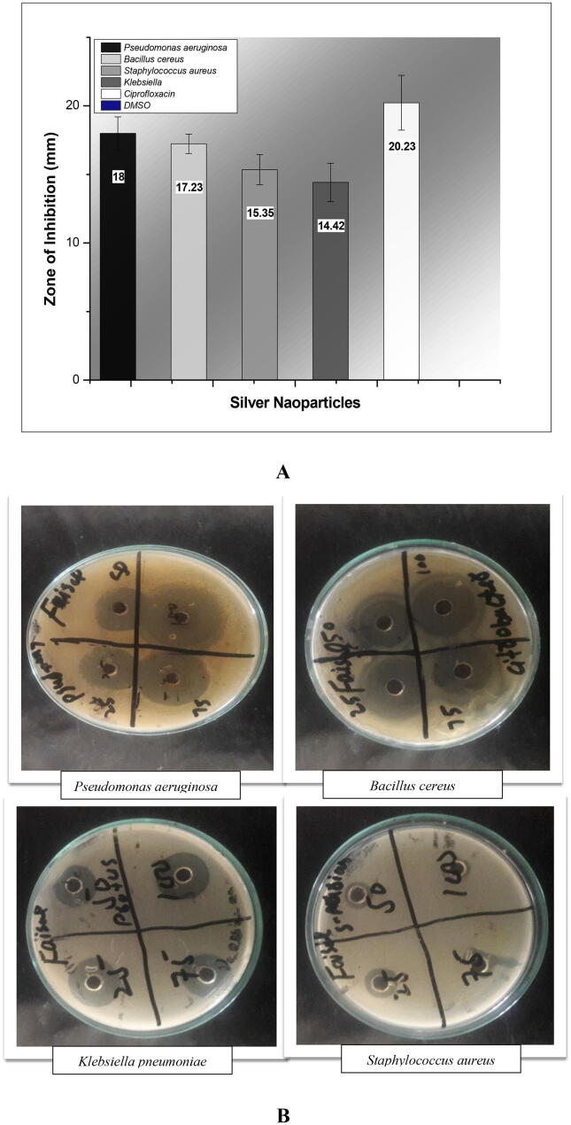
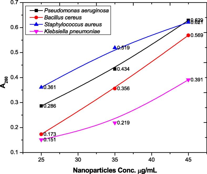
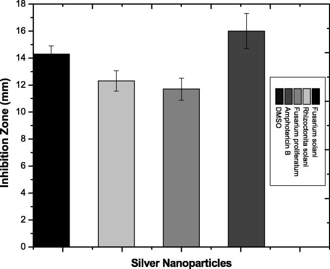
In recent years green nanotechnology gained significant importance to synthesize nanoparticles due to their cost effectiveness and biosafety. In the current study, silver nanoparticles were synthesized by using extract of Spirogyra hyalina as a capping and reducing agent. The synthesized nanoparticles were characterized by UV-Visible spectroscopy, Fourier transform infrared spectroscopy, Scanning electron microscopy, energy dispersive X-ray spectroscopy, and X-ray diffractive analysis. Silver nanoparticles give a characteristic Surface Plasmon Resonance peak of 451 nm at 2.21 a.u (arbitrary unit). SEM micrograph revealed the spherical morphology and average grain size of 52.7 nm. Furthermore, antibacterial, antifungal, insecticidal, antioxidant and membrane damage activities were determined. The maximum antibacterial and antifungal activity was observed for Pseudomonas aeruginosa (18 ± 1.2 mm) and Fusarium solani (14.3 ± 0.6 mm), respectively. In membrane damage assay, Pseudomonas aeruginosa absorbed A260 wavelength and gave maximum peak values of 0.286, 0.434 and 0.629 at 25, 35 and 45 µg/mL of silver nanoparticles. The membrane damage assay confirmed that nanoparticles are involved in bacterial cell membrane damage. At 500 ppm silver nanoparticles showed 30% mortality against Tribolium castaneum (a common grain pest). The silver nanoparticles also showed potent antioxidant activity and successfully scavenged the DPPH free radicals upto 53.43 ± 0.17, 43.26 ± 0.97, 31.39 ± 0.33, 24.62 ± 0.85, and 14.13 ± 0.12% at a concentration of 400, 200, 100, 50, and 25 µg/mL of nanoparticles, respectively. It is concluded that silver nanoparticles can easily be synthesized by using green algae Spirogyra hyalina as a capping and reducing agent. Silver nanoparticles showed potent biomedical activities and thus can be used for therapeutic applications invitro and invivo.
Keywords: Bio catalytic efficacy; Biosynthesis; Nanoparticles; Silver; Spirogyra.
© 2021 The Author(s).
Conflict of interest statement
The authors declare that they have no known competing financial interests or personal relationships that could have appeared to influence the work reported in this paper.
Figures

References
-
- Abdel-Raouf N., Alharbi R.M., Al-Enazi N.M., Alkhulaifi M.M., Ibraheem I.B.M. Rapid biosynthesis of silver nanoparticles using the marine red alga Laurencia catarinensis and their characterization. Beni-Suef University J. Basic Appl. Sci. 2018;7(1):150–157.
-
- Al-Radadi N.S. Artichoke (Cynara scolymus L.,) mediated rapid analysis of silver nanoparticles and their utilisation on the cancer cell treatments. Journal of Computational and Theoretical Nanoscience. 2018;15(6–7):1818–1829. doi: 10.1166/jctn.2018.7317. - DOI
-
- Al-Radadi N.S. Green synthesis of platinum nanoparticles using Saudi’s Dates extract and their usage on the cancer cell treatment. Arabian Journal of Chemistry. 2019;12(3):330–349. doi: 10.1016/j.arabjc.2018.05.008. - DOI
-
- Al-Radadi, N. S. 2021. Facile one-step green synthesis of gold nanoparticles (AuNp) using Licorice root extract: antimicrobial and anticancer study against HepG2 cell line Arab. J.Chem 14 (2021)1-25. 10.1016/j.arabjc.2020.102956 - DOI
-
- Al-Radadi, N. S. 2021. Green Biosynthesis of Flaxseed Gold Nanoparticles (Au-NPs) as Potent Anticancer Agent Against Breast Cancer Cells. J.Saudi.Chem.Society 25(6) (2021)1-22. 10.1016/j.jscs.2021.101243 - DOI
LinkOut - more resources
Full Text Sources
