

Molecular Evaluation of Endometrial Dedifferentiated Carcinoma, Endometrioid Carcinoma, Carcinosarcoma, and Serous Carcinoma Using a Custom-Made Small Cancer Panel
- PMID: 35002543
- PMCID: PMC8734147
- DOI: 10.3389/pore.2021.1610013
Molecular Evaluation of Endometrial Dedifferentiated Carcinoma, Endometrioid Carcinoma, Carcinosarcoma, and Serous Carcinoma Using a Custom-Made Small Cancer Panel
Abstract
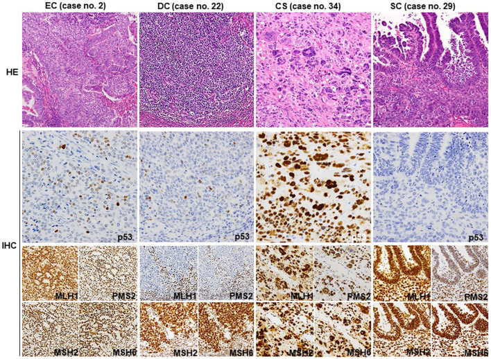
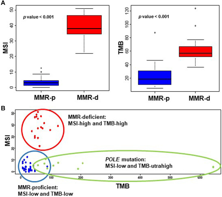
It is often difficult to histologically differentiate among endometrial dedifferentiated carcinoma (DC), endometrioid carcinoma (EC), serous carcinoma (SC), and carcinosarcoma (CS) due to the presence of solid components. In this study, we aimed to categorize these carcinomas according to The Cancer Genome Atlas (TCGA) classification using a small custom-made cancer genome panel (56 genes and 17 microsatellite regions) for integrated molecular diagnosis. A total of 36 endometrial cancer cases with solid components were assessed using IHC, next-generation sequencing (NGS), and the custom-made panel. Among 19 EC cases, six were categorized as MMR-deficient (MMR-d) and eight were classified as having a nonspecific molecular profile. Three EC cases were classified as POLE mutation (POLEmut)-type, which had a very high tumor mutation burden (TMB) and low microsatellite instability (MSI). Increased TMB and MSI were observed in all three DC cases, classified as MMR-d with mutations in MLH1 and POLD1. Except for one case classified as MMR-d, all SC cases exhibited TP53 mutations and were classified as p53 mutation-type. SC cases also exhibited amplification of CCND1, CCNE1, and MYC. CS cases were classified as three TCGA types other than the POLEmut-type. The IHC results for p53 and ARID1A were almost consistent with their mutation status. NGS analysis using a small panel enables categorization of endometrial cancers with solid proliferation according to TCGA classification. As TCGA molecular classification does not consider histological findings, an integrated analytical procedure including IHC and NGS may be a practical diagnostic tool for endometrial cancers.
Keywords: endometrial cancers; integrated molecular diagnosis; microsatellite instability; solid proliferation; tumor mutation burden.
Copyright © 2021 Kobayashi, Kitazono, Akahane, Yanazume, Kamio, Togami, Nohara, Sakamoto, Yokoyama, Tabata, Kobayashi and Tanimoto.
Conflict of interest statement
SN and IS are employed by Mitsubishi Space Software. The remaining authors declare that the research was conducted in the absence of any commercial or financial relationships that could be construed as a potential conflict of interest.
Figures
Similar articles
-
Utility of a custom designed next generation DNA sequencing gene panel to molecularly classify endometrial cancers according to The Cancer Genome Atlas subgroups.BMC Med Genomics. 2020 Nov 30;13(1):179. doi: 10.1186/s12920-020-00824-8. BMC Med Genomics. 2020. PMID: 33256706 Free PMC article.
-
Analysis of 108 patients with endometrial carcinoma using the PROMISE classification and additional genetic analyses for MMR-D.Gynecol Oncol. 2020 Apr;157(1):245-251. doi: 10.1016/j.ygyno.2020.01.019. Epub 2020 Jan 21. Gynecol Oncol. 2020. PMID: 31980219
-
Use of mutation profiles to refine the classification of endometrial carcinomas.J Pathol. 2012 Sep;228(1):20-30. doi: 10.1002/path.4056. Epub 2012 Jul 18. J Pathol. 2012. PMID: 22653804 Free PMC article.
-
TCGA Classification of Endometrial Cancer: the Place of Carcinosarcoma.Pathol Oncol Res. 2020 Oct;26(4):2067-2073. doi: 10.1007/s12253-020-00829-9. Epub 2020 May 29. Pathol Oncol Res. 2020. PMID: 32472441
-
Molecular Genetics of Endometrial Carcinoma.Annu Rev Pathol. 2019 Jan 24;14:339-367. doi: 10.1146/annurev-pathol-020117-043609. Epub 2018 Oct 17. Annu Rev Pathol. 2019. PMID: 30332563 Review.
Cited by
-
Abnormal p53 High-Grade Endometrioid Endometrial Cancer: A Systematic Review and Meta-Analysis.Cancers (Basel). 2024 Dec 26;17(1):38. doi: 10.3390/cancers17010038. Cancers (Basel). 2024. PMID: 39796669 Free PMC article. Review.
-
The Advantages of Next-Generation Sequencing Molecular Classification in Endometrial Cancer Diagnosis.J Clin Med. 2023 Nov 22;12(23):7236. doi: 10.3390/jcm12237236. J Clin Med. 2023. PMID: 38068290 Free PMC article.
-
Cervical Cytology Preserves Histologically Detected Surface Epithelial Slackening, Unique to the POLE Mutation-subtype in Endometrial Cancer.In Vivo. 2024 Jan-Feb;38(1):321-333. doi: 10.21873/invivo.13442. In Vivo. 2024. PMID: 38148087 Free PMC article.
-
Prospects of POLD1 in Human Cancers: A Review.Cancers (Basel). 2023 Mar 22;15(6):1905. doi: 10.3390/cancers15061905. Cancers (Basel). 2023. PMID: 36980791 Free PMC article. Review.
-
Uterine Carcinosarcoma (UCS): A Literature Review and Survival Analysis from a Retrospective Cohort Study.Cancers (Basel). 2024 Nov 21;16(23):3905. doi: 10.3390/cancers16233905. Cancers (Basel). 2024. PMID: 39682097 Free PMC article.
References
-
- Kim K-R, Lax SF, Lazar AJ, Longacre TA, Malpica A, Matias-Guiu X, et al. Tumours of Uterine Corpus. In: The WHO Classification of Tumours Editorial boardFemale Genital Tumours. 5th ed. Lyon: IARC; (2020). p. 245–308.
MeSH terms
LinkOut - more resources
Full Text Sources
Research Materials
Miscellaneous