

Association Between 18-FDG Positron Emission Tomography and MRI Biomarkers of Plaque Vulnerability in Patients With Symptomatic Carotid Stenosis
- PMID: 35002912
- PMCID: PMC8732361
- DOI: 10.3389/fneur.2021.731744
Association Between 18-FDG Positron Emission Tomography and MRI Biomarkers of Plaque Vulnerability in Patients With Symptomatic Carotid Stenosis
Abstract
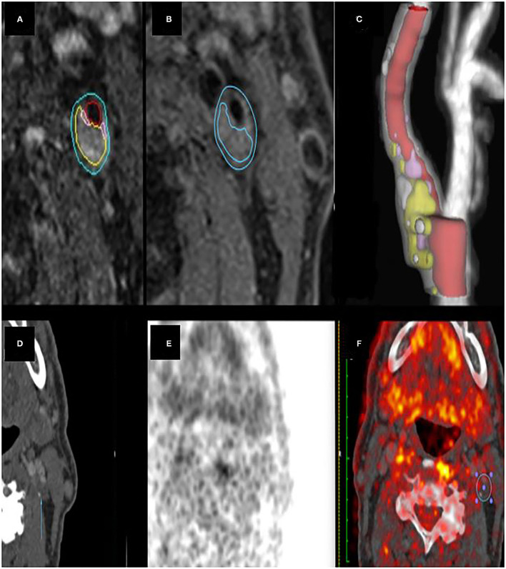
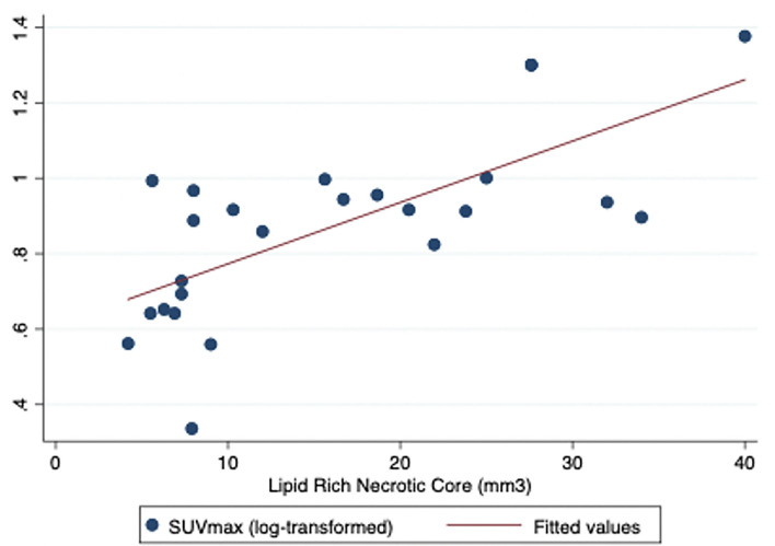
Purpose: Pathologic studies suggest that unstable plaque morphology and inflammation are associated with cerebrovascular events. 18F-fluorodeoxyglucose positron emission tomography (18FDG-PET) is a validated technique for non-invasive imaging of inflammation-related plaque metabolism, and MRI can identify morphologic features of plaque instability. The aim of this study was to investigate the association of selected imaging characteristics of plaque vulnerability measured with MRI and PET in patients with symptomatic carotid stenosis. Methods: Patients from the BIOVASC study were selected based on the following inclusion criteria: (1) age ≥ 50 years; (2) recent (<30 days) ischaemic stroke (modified Rankin scale ≤3) or motor/speech/vision TIA; (3) ipsilateral internal carotid artery stenosis (≥5 0% lumen-narrowing); (4) carotid PET/CTA and MRI completed. Semi-automated plaque analysis of MRI images was performed to quantify morphologic features of plaque instability. PET images were co-registered with CTA and inflammation-related metabolism expressed as maximum standardised uptake value (SUVmax). Results: Twenty-five patients met inclusion criteria (72% men, mean age 65 years). MRI-measured plaque volume was greater in men (1,708-1,286 mm3, p = 0.03), patients who qualified with stroke (1,856-1,440 mm3, p = 0.05), and non-statin users (1,325-1,797 mm3, p = 0.03). SUVmax was associated with MRI-measured plaque lipid-rich necrotic core (LRNC) in the corresponding axial slice (r s = 0.64, p < 0.001) and was inversely associated with whole-plaque fibrous cap thickness (r s = -0.4, p = 0.02) and calcium volume (r s = -0.4, p = 0.03). Conclusion: This study demonstrated novel correlations of non-invasive imaging biomarkers of inflammation-related plaque metabolism with morphological MRI markers of plaque instability. If replicated, our findings may support the application of combined MRI and PET to detect vulnerable plaque in future clinical practise and randomised trials.
Keywords: MRI; PET; atherosclerosis; carotid; plaque inflammation; plaque segmentation; vulnerable plaque biomarker.
Copyright © 2021 Giannotti, McNulty, Foley, McCabe, Barry, Crowe, Dolan, Harbison, Horgan, Kavanagh, O'Connell, Marnane, Murphy, Donnell, O'Donohoe, Williams and Kelly.
Conflict of interest statement
The authors declare that the research was conducted in the absence of any commercial or financial relationships that could be construed as a potential conflict of interest.
Figures
Similar articles
-
Cohort profile: BIOVASC-late, a prospective multicentred study of imaging and blood biomarkers of carotid plaque inflammation and risk of late vascular recurrence after non-severe stroke in Ireland.BMJ Open. 2020 Jul 19;10(7):e038607. doi: 10.1136/bmjopen-2020-038607. BMJ Open. 2020. PMID: 32690537 Free PMC article.
-
Contemporary carotid imaging: from degree of stenosis to plaque vulnerability.J Neurosurg. 2016 Jan;124(1):27-42. doi: 10.3171/2015.1.JNS142452. Epub 2015 Jul 31. J Neurosurg. 2016. PMID: 26230478 Review.
-
Carotid Plaque Inflammation Imaged by 18F-Fluorodeoxyglucose Positron Emission Tomography and Risk of Early Recurrent Stroke.Stroke. 2019 Jul;50(7):1766-1773. doi: 10.1161/STROKEAHA.119.025422. Epub 2019 Jun 6. Stroke. 2019. PMID: 31167623 Clinical Trial.
-
Carotid Plaque Inflammation Imaged by PET and Prediction of Recurrent Stroke at 5 Years.Neurology. 2021 Dec 7;97(23):e2282-e2291. doi: 10.1212/WNL.0000000000012909. Epub 2021 Oct 5. Neurology. 2021. PMID: 34610991
-
MR imaging of vulnerable carotid plaque.Cardiovasc Diagn Ther. 2020 Aug;10(4):1019-1031. doi: 10.21037/cdt.2020.03.12. Cardiovasc Diagn Ther. 2020. PMID: 32968658 Free PMC article. Review.
Cited by
-
Positron emission tomography and its role in the assessment of vulnerable plaques in comparison to other imaging modalities.Front Med (Lausanne). 2024 Feb 15;10:1293848. doi: 10.3389/fmed.2023.1293848. eCollection 2023. Front Med (Lausanne). 2024. PMID: 38425695 Free PMC article. Review.
-
Diagnostic potential of TSH to HDL cholesterol ratio in vulnerable carotid plaque identification.Front Cardiovasc Med. 2024 May 28;11:1333908. doi: 10.3389/fcvm.2024.1333908. eCollection 2024. Front Cardiovasc Med. 2024. PMID: 38863898 Free PMC article.
-
MRI ensemble model of plaque and perivascular adipose tissue as PET-equivalent for identifying carotid atherosclerotic inflammation.EJNMMI Res. 2025 Aug 6;15(1):103. doi: 10.1186/s13550-025-01293-9. EJNMMI Res. 2025. PMID: 40768105 Free PMC article.
-
Machine learning using scRNA-seq Combined with bulk-seq to identify lactylation-related hub genes in carotid arteriosclerosis.Sci Rep. 2025 May 22;15(1):17794. doi: 10.1038/s41598-025-00834-5. Sci Rep. 2025. PMID: 40404675 Free PMC article.
-
Role of plaque inflammation in symptomatic carotid stenosis.Front Neurol. 2023 Jan 24;14:1086465. doi: 10.3389/fneur.2023.1086465. eCollection 2023. Front Neurol. 2023. PMID: 36761341 Free PMC article.
References
-
- Saba L, Yuan C, Hatsukami TS, Balu N, Qiao Y, DeMarco JK, et al. . Vessel wall imaging study group of the american society of neuroradiology. carotid artery wall imaging: perspective and guidelines from the asnr vessel wall imaging study group and expert consensus recommendations of the American Society of Neuroradiology. Am J Neuroradiol. (2018) 39:E9–E31. 10.3174/ajnr.A5488 - DOI - PMC - PubMed
LinkOut - more resources
Full Text Sources
Miscellaneous