

A Preliminary DTI Tractography Study of Developmental Neuroplasticity 5-15 Years After Early Childhood Traumatic Brain Injury
- PMID: 35002913
- PMCID: PMC8732947
- DOI: 10.3389/fneur.2021.734055
A Preliminary DTI Tractography Study of Developmental Neuroplasticity 5-15 Years After Early Childhood Traumatic Brain Injury
Abstract
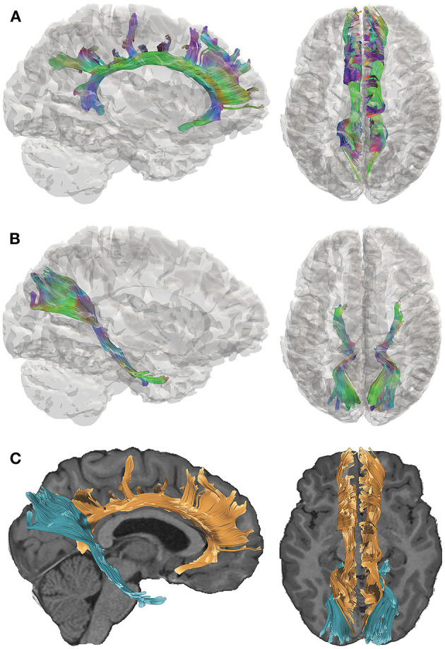
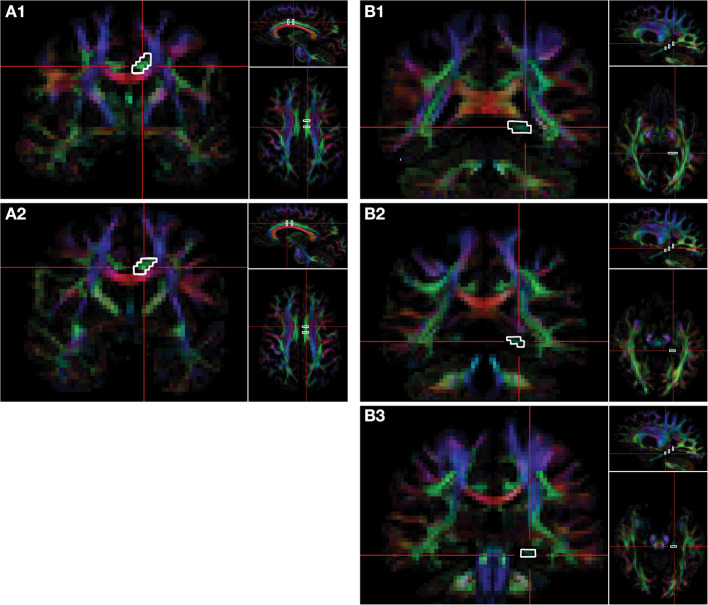
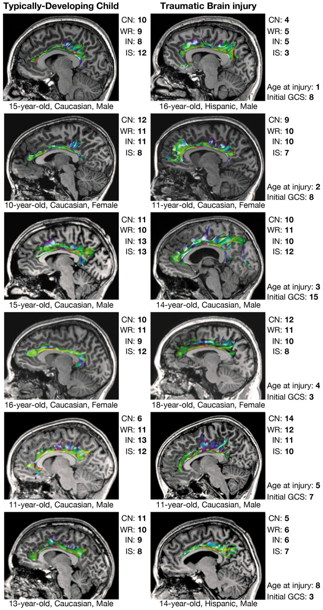
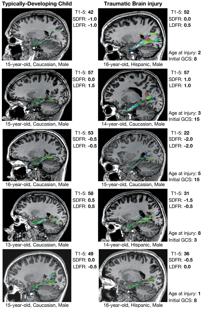
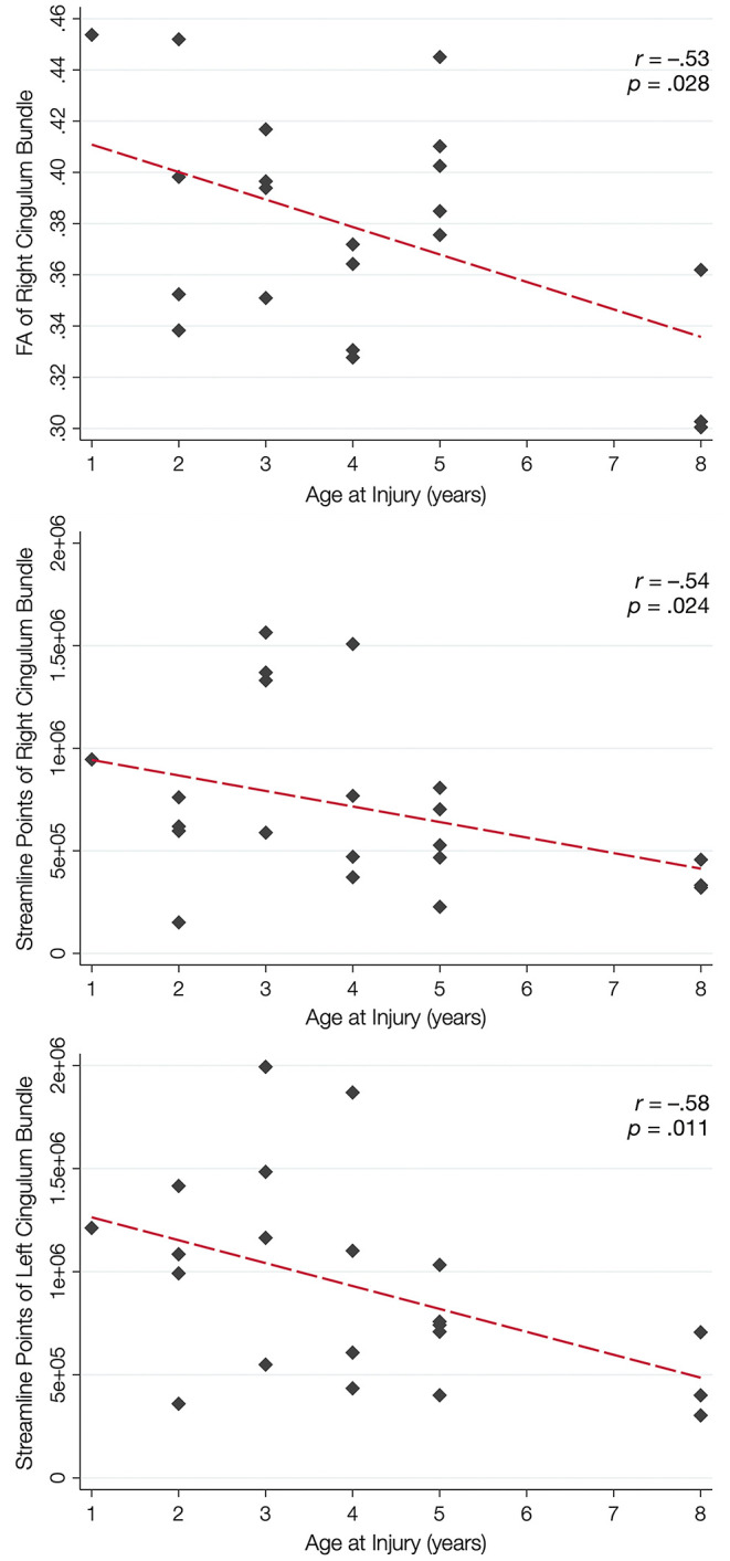
Plasticity is often implicated as a reparative mechanism when addressing structural and functional brain development in young children following traumatic brain injury (TBI); however, conventional imaging methods may not capture the complexities of post-trauma development. The present study examined the cingulum bundles and perforant pathways using diffusion tensor imaging (DTI) in 21 children and adolescents (ages 10-18 years) 5-15 years after sustaining early childhood TBI in comparison with 19 demographically-matched typically-developing children. Verbal memory and executive functioning were also evaluated and analyzed in relation to DTI metrics. Beyond the expected direction of quantitative DTI metrics in the TBI group, we also found qualitative differences in the streamline density of both pathways generated from DTI tractography in over half of those with early TBI. These children exhibited hypertrophic cingulum bundles relative to the comparison group, and the number of tract streamlines negatively correlated with age at injury, particularly in the late-developing anterior regions of the cingulum; however, streamline density did not relate to executive functioning. Although streamline density of the perforant pathway was not related to age at injury, streamline density of the left perforant pathway was significantly and positively related to verbal memory scores in those with TBI, and a moderate effect size was found in the right hemisphere. DTI tractography may provide insight into developmental plasticity in children post-injury. While traditional DTI metrics demonstrate expected relations to cognitive performance in group-based analyses, altered growth is reflected in the white matter structures themselves in some children several years post-injury. Whether this plasticity is adaptive or maladaptive, and whether the alterations are structure-specific, warrants further investigation.
Keywords: brain development; diffusion tensor imaging; neuroplasticity; pediatric traumatic brain injury; structural neuroimaging; tractography.
Copyright © 2021 Wilde, Hyseni, Lindsey, Faber, McHenry, Bigler, Biekman, Hollowell, McCauley, Hunter, Ewing-Cobbs, Aitken, MacLeod, Chu, Noble-Haeusslein and Levin.
Conflict of interest statement
The authors declare that the research was conducted in the absence of any commercial or financial relationships that could be construed as a potential conflict of interest.
Figures
Similar articles
-
Acute pediatric traumatic brain injury severity predicts long-term verbal memory performance through suppression by white matter integrity on diffusion tensor imaging.Brain Imaging Behav. 2020 Oct;14(5):1626-1637. doi: 10.1007/s11682-019-00093-9. Brain Imaging Behav. 2020. PMID: 31134584 Free PMC article.
-
Ten-year outcome of early childhood traumatic brain injury: Diffusion tensor imaging of the ventral striatum in relation to executive functioning.Brain Inj. 2016;30(13-14):1635-1641. doi: 10.1080/02699052.2016.1199910. Epub 2016 Sep 28. Brain Inj. 2016. PMID: 27680309
-
Evaluating the relationship between memory functioning and cingulum bundles in acute mild traumatic brain injury using diffusion tensor imaging.J Neurotrauma. 2010 Feb;27(2):303-7. doi: 10.1089/neu.2009.1110. J Neurotrauma. 2010. PMID: 19877826 Free PMC article.
-
Diffusion tensor imaging (DTI) findings in adult civilian, military, and sport-related mild traumatic brain injury (mTBI): a systematic critical review.Brain Imaging Behav. 2018 Apr;12(2):585-612. doi: 10.1007/s11682-017-9708-9. Brain Imaging Behav. 2018. PMID: 28337734
-
Traumatic brain injury, major depression, and diffusion tensor imaging: making connections.Brain Res Rev. 2010 Sep;64(1):213-40. doi: 10.1016/j.brainresrev.2010.04.003. Epub 2010 Apr 11. Brain Res Rev. 2010. PMID: 20388528 Review.
Cited by
-
Neuroimaging Correlates of Functional Outcome Following Pediatric TBI.Adv Neurobiol. 2024;42:33-84. doi: 10.1007/978-3-031-69832-3_3. Adv Neurobiol. 2024. PMID: 39432037 Review.
-
Early adversity causes sex-specific deficits in perforant pathway connectivity and contextual memory in adolescent mice.Biol Sex Differ. 2024 May 7;15(1):39. doi: 10.1186/s13293-024-00616-0. Biol Sex Differ. 2024. PMID: 38715106 Free PMC article.
-
Learning by Heart or with Heart: Brain Asymmetry Reflects Pedagogical Practices.Brain Sci. 2023 Aug 31;13(9):1270. doi: 10.3390/brainsci13091270. Brain Sci. 2023. PMID: 37759871 Free PMC article.
-
Symptom Persistence Relates to Volume and Asymmetry of the Limbic System after Mild Traumatic Brain Injury.J Clin Med. 2024 Aug 30;13(17):5154. doi: 10.3390/jcm13175154. J Clin Med. 2024. PMID: 39274367 Free PMC article.
-
The impact of loneliness and social adaptation on depressive symptoms: Behavioral and brain measures evidence from a brain health perspective.Front Psychol. 2023 Mar 13;14:1096178. doi: 10.3389/fpsyg.2023.1096178. eCollection 2023. Front Psychol. 2023. PMID: 37077845 Free PMC article.
References
LinkOut - more resources
Full Text Sources