. 2021 Dec 23:12:774490.
doi: 10.3389/fmicb.2021.774490. eCollection 2021.

Optimized Attenuated Salmonella Typhimurium Suppressed Tumor Growth and Improved Survival in Mice

Affiliations

Optimized Attenuated Salmonella Typhimurium Suppressed Tumor Growth and Improved Survival in Mice

Kang Liang et al. Front Microbiol. .

Abstract

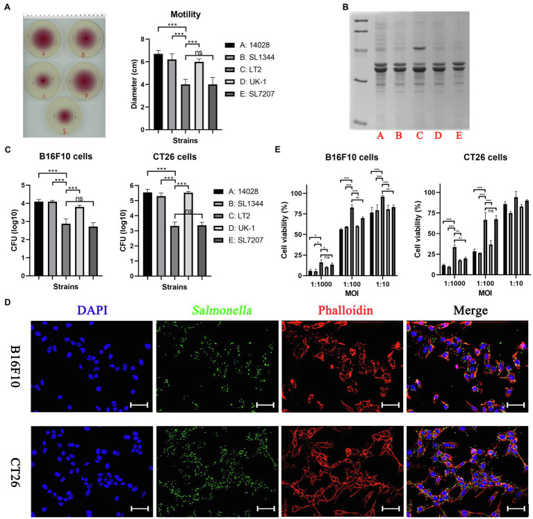
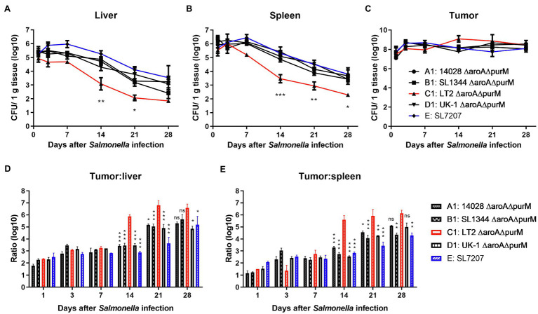
The gram-negative facultative anaerobic bacteria Salmonella enterica serovar Typhimurium (hereafter S. Typhimurium) has always been considered as one candidate of anti-tumor agents or vectors for delivering drug molecules. In this study, we compared several widely studied S. Typhimurium strains in their anti-tumor properties aiming to screen out the best one for further optimization and use in cancer therapy. In terms of the motility, virulence and anti-tumor efficacy, the three strains 14028, SL1344, and UK-1 were similar and obviously better than LT-2, and UK-1 showed the best phenotypes among them. Therefore, the strain UK-1 (D) was selected for the following studies. Its auxotrophic mutant strain (D1) harboring ∆aroA and ∆purM mutations was further optimized through the modification of lipid A structure, generating a new strain named D2 with stronger immunostimulatory activity. Finally, the ∆asd derivative of D2 was utilized as one live vector to deliver anti-tumor molecules including the angiogenesis inhibitor endostatin and apoptosis inducer TRAIL and the therapeutic and toxic-side effects were evaluated in mouse models of colon carcinoma and melanoma. After intraperitoneal infection, engineered Salmonella bacteria equipped with endostatin and/or TRAIL significantly suppressed the tumor growth and prolonged survival of tumor-bearing mice compared to PBS or bacteria carrying the empty plasmid. Consistently, immunohistochemical studies confirmed the colonization of Salmonella bacteria and the expression of anti-tumor molecules inside tumor tissue, which were accompanied by the increase of cell apoptosis and suppression of tumor angiogenesis. These results demonstrated that the beneficial anti-tumor efficacy of attenuated S. Typhimurium bacteria could be improved through delivery of drug molecules with powerful anti-tumor activities.

Keywords: Salmonella Typhimurium; TRAIL; cancer therapy; delivery; endostatin.


Conflict of interest statement

The authors declare that the research was conducted in the absence of any commercial or financial relationships that could be construed as a potential conflict of interest.

Figures

Figure 1
Figure 1
The in vitro phenotypes of different Salmonella Typhimurium strains. (A) Swimming motility of bacterial strains was tested on 0.3% semi-solid agar plates. The swimming diameter was measured with a ruler. (B) Outer membrane proteins purified from different Salmonella strains were stained by coomassie brilliant blue. (C) The ability of bacterial strains to invade cancer cells was determined by counting alive bacterial cells (CFU). Briefly, cancer cells were infected by Salmonella for 2 h at an MOI of 100:1, and further cultured with medium containing gentamycin for 1 h to kill extracellular bacteria. (D) The invasion of Salmonella into cancer cells was also confirmed by the immunofluorescence assay. For visualization by fluorescence microscopy, Salmonella bacteria (green) were stained using rabbit anti-Salmonella primary antibody and anti-rabbit secondary antibody conjugated with Alexa Fluor 488, followed by cell nuclei (blue) and actin (red) stained by DAPI and phalloidin, respectively. Scale bar, 50 μm. (E) The toxicity of bacteria to cancer cells after 6 h of co-incubation was assessed by the CCK-8 assay. The significance of differences among groups were analyzed by two-way ANOVA analysis followed by Tukey’s multiple comparisons test and indicated by asterisks (*p < 0.05; **p < 0.01; and ***p < 0.001).
Figure 2
Figure 2
The in vivo colonization of different S. Typhimurium auxotrophic strains in tumor-bearing mice. To determinate the in vivo colonization of different S. Typhimurium auxotrophic strains in tumor-bearing mice, four tumor-bearing mice were euthanized at each indicated dpi, and tumor and normal tissues were taken sterilely. Bacterial burdens in the liver (A), spleen (B), and tumor tissues (C) were measured by plating serial dilutions of tissue homogenates and expressed as CFU g-1 tissue. (D,E) Tumor-to-normal tissue ratios at different dpi were calculated. At dpi 14, 21, and 28, the significance of the difference between the colonization number of C1 and other strains was analyzed. The colonization profiles of C1 and other strains were compared and difference significance was analyzed through two-way ANOVA analysis followed by Tukey’s multiple comparisons test (*p < 0.05; **p < 0.01; and ***p < 0.001).
Figure 3
Figure 3
The anti-tumor efficacy of different S. Typhimurium auxotrophic strains in mouse models. Tumor-bearing mice were randomly divided into groups of eight mice each and intraperitoneally injected with 100 μl of PBS or different S. Typhimurium auxotrophic strains (5 × 106 CFU). Tumor volume of CT26 colon carcinoma- (A) and B16F10 melanoma-bearing mice (B) were measured every 2–3 days and difference significance between PBS and other groups was analyzed through two-way ANOVA analysis followed by Tukey’s multiple comparisons test (*p < 0.05; **p < 0.01; and ***p < 0.001). (C) The Kaplan–Meier survival curves for mice bearing B16F10 melanoma were monitored. (D) The median survival time was also analyzed and shown as the median with upper and lower limits. (E,F) During animal experiments, body weight of tumor-bearing mice was measured as an indicator for general health status.
Figure 4
Figure 4
The expression of endostatin and/or TRAIL by optimized Salmonella and their in vitro cell-killing activities. (A,B) The expression of endostatin (ES, RGD4C-ES, RGD10-ES, and PSMA-ES; 25.2, 28.1, 28.4, and 52.6 kDa) and TRAIL (34.3 kDa) by engineered Salmonella strain D2-asd was detected by western blotting, respectively. The samples tested were prepared from bacterial lysates. (C) Recombinant Salmonella co-expressing endostatin and TRAIL were also constructed and verified in the same way. (D–F) The toxicity of recombinant Salmonella bacteria expressing endostatin and/or TRAIL to HUVECs and cancer cells at the MOI of 1:100 or 1:10 were tested by the CCK-8 assay. Besides, the filtered medium supernatant of bacterial culture containing secreted anti-tumor molecules was also added to cells for testing. The cell viability shown (%) was relative to that of untreated cells, followed by comparison among groups by two-way ANOVA analysis. The cell viability shown (%) was relative to that of untreated cells. Difference significance between ST/3342 and other groups was analyzed through two-way ANOVA analysis followed by Tukey’s multiple comparisons test (*p < 0.05; **p < 0.01; and ***p < 0.001).
Figure 5
Figure 5
The in vivo anti-tumor efficacy of engineered Salmonella expressing endostatin. Tumor-bearing mice were randomly divided into groups of eight mice each and intraperitoneally injected with 100 μl of PBS or 5 × 106 CFU of attenuated Salmonella (D2-asd) carrying pYA3342 or plasmids expressing ES, RGD4C-ES, RGD10-ES, and PSMA-ES, respectively. (A,B) Tumor volume of mice from different groups were measured every 2–3 days and difference significance between PBS and other groups was subjected to multiple comparisons through two-way ANOVA analysis (*p < 0.05; **p < 0.01; and ***p < 0.001). (C) for mice bearing melanoma were monitored and the median survival time (D) was shown as the median with upper and lower limits. (E,F) Meanwhile, body weight of tumor-bearing mice was recorded.
Figure 6
Figure 6
The in vivo anti-tumor efficacy of engineered Salmonella bacteria expressing TRAIL and endostatin. Tumor-bearing mice were randomly divided into four groups and intraperitoneally injected with 100 μl of PBS or 5 × 106 CFU of attenuated Salmonella bacteria (D2-asd) carrying empty pYA3342, TRAIL-expression and TRAIL-RGD4C-ES co-expression plasmids, respectively. (A,B) The change of tumor volume were recorded every 2–3 days and difference significance between PBS and other groups was analyzed through two-way ANOVA analysis followed by Tukey’s multiple comparisons test (*p < 0.05; **p < 0.01; and ***p < 0.001). (C) The Kaplan–Meier survival curves for mice bearing melanoma were monitored and difference significance was analyzed by the log-rank test. (D) The median survival time of mice in different groups was shown as the median with upper and lower limits. (E,F) Body weight of tumor-bearing mice was also recorded during animal experiments.
Figure 7
Figure 7
Immunohistochemical studies on tumor tissue. Two weeks after tumor-bearing mice received different treatments, tumor samples were taken for immunohistochemical studies as described above. The accumulation of Salmonella bacteria and the expression of anti-tumor molecules inside tumor tissue were detected using specific primary antibodies, respectively, against Salmonella, endostatin and TRAIL. Tumor microvascular density (MVD) and cell apoptosis level were also evaluated by staining for CD34 and cleaved caspase-3, respectively. Scale bar, 50 μm.
Figure 8
Figure 8
Pathological studies on normal tissues. Standard H.E staining was performed on the liver and spleen tissues taken during animal experiments. In Salmonella infection groups, disorderly arrangement of hepatic cords accompanied by swelling and degeneration of hepatocytes was observed in the liver, and the spleen appeared slight inflammation and the infiltration of phagocytic cells and inflammatory cells. There were no obvious pathological changes in PBS treatment group.

Similar articles

Cited by

References

    1. Afshar-Oromieh A., Babich J. W., Kratochwil C., Giesel F. L., Eisenhut M., Kopka K., et al. . (2016). The rise of PSMA ligands for diagnosis and therapy of prostate cancer. J. Nucl. Med. 57(Suppl. 3), 79S–89S. doi: 10.2967/jnumed.115.170720, PMID: - DOI - PubMed
    1. Arrach N., Cheng P., Zhao M., Santiviago C. A., Hoffman R. M., McClelland M. (2010). High-throughput screening for Salmonella avirulent mutants that retain targeting of solid tumors. Cancer Res. 70, 2165–2170. doi: 10.1158/0008-5472.CAN-09-4005, PMID: - DOI - PMC - PubMed
    1. Curtiss R., Hassan J. O. (1996). Nonrecombinant and recombinant avirulent Salmonella vaccines for poultry. Vet. Immunol. Immunopathol. 54, 365–372. doi: 10.1016/s0165-2427(96)05683-8, PMID: - DOI - PubMed
    1. Danhier F., Le Breton A., Preat V. (2012). RGD-based strategies to target αvβ3 integrin in cancer therapy and diagnosis. Mol. Pharm. 9, 2961–2973. doi: 10.1021/mp3002733, PMID: - DOI - PubMed
    1. Daniels R. A., Turley H., Kimberley F. C., Liu X. S., Mongkolsapaya J., Ch'En P., et al. . (2005). Expression of TRAIL and TRAIL receptors in normal and malignant tissues. Cell Res. 15, 430–438. doi: 10.1038/sj.cr.7290311, PMID: - DOI - PubMed

LinkOut - more resources