. 2021 Dec 24:12:760770.
doi: 10.3389/fimmu.2021.760770. eCollection 2021.

Physical Interactions With Bacteria and Protozoan Parasites Establish the Scavenger Receptor SSC4D as a Broad-Spectrum Pattern Recognition Receptor

Affiliations

Physical Interactions With Bacteria and Protozoan Parasites Establish the Scavenger Receptor SSC4D as a Broad-Spectrum Pattern Recognition Receptor

Marcos S Cardoso et al. Front Immunol. .

Abstract

Since the pioneering discoveries, by the Nobel laureates Jules Hoffmann and Bruce Beutler, that Toll and Toll-like receptors can sense pathogenic microorganisms and initiate, in vertebrates and invertebrates, innate immune responses against microbial infections, many other families of pattern recognition receptors (PRRs) have been described. One of such receptor clusters is composed by, if not all, at least several members of the scavenger receptor cysteine-rich (SRCR) superfamily. Many SRCR proteins are plasma membrane receptors of immune cells; however, a small subset consists of secreted receptors that are therefore in circulation. We here describe the first characterization of biological and functional roles of the circulating human protein SSC4D, one of the least scrutinized members of the family. Within leukocyte populations, SSC4D was found to be expressed by monocytes/macrophages, neutrophils, and B cells, but its production was particularly evident in epithelial cells of several organs and tissues, namely, in the kidney, thyroid, lung, placenta, intestinal tract, and liver. Similar to other SRCR proteins, SSC4D shows the capacity of physically binding to different species of bacteria, and this opsonization can increase the phagocytic capacity of monocytes. Importantly, we have uncovered the capacity of SSC4D of binding to several protozoan parasites, a singular feature seldom described for PRRs in general and here demonstrated for the first time for an SRCR family member. Overall, our study is pioneer in assigning a PRR role to SSC4D.

Keywords: bacteria; circulating receptors; parasites; pattern recognition receptors; scavenger receptor cysteine-rich.


Conflict of interest statement

The authors declare that the research was conducted in the absence of any commercial or financial relationships that could be construed as a potential conflict of interest.

Figures

Figure 1
Figure 1
Amino acid sequence and structure of group B scavenger receptor cysteine-rich (SRCR) domains of SSC4D, CD5L, and CD6. (A) SRCR domains are typically ~100–110 amino acids in length compacted into a heart-shaped fold, where a six/seven-stranded β sheet cradles a core α1-helix. Each line represents one SRCR domain of the indicated protein. Amino acid sequences were aligned using Clustal Omega and MView (40). Amino acid side chain color codes for conserved residues: Green/black, aliphatic; Green/white, aromatic; Blue, anionic; Red, cationic; Cyan, polar; Magenta, amide; Yellow, sulfur-containing. Intrachain disulfide bonds established between conserved cysteine residues are shown on the top by connecting lines. (B) Schematic representation of the protein structures of SSC4D-FL, SSC4D-d1d2, SSC4D-d3d4, CD5L, and extracellular domain of CD6 (sCD6). SRCR domains are represented as dark cylinders. Putative O-linked glycosylation sites are represented as short vertical lines and N-linked glycosylation sites as lines topped with red circles. N and C termini of the proteins are indicated by “N” and “C,” respectively. Design was created using BioRender.com. (C) SSC4D protein expression detected by western blotting from cell lysates of Hep G2, Caco-2, K562, and HeLa cells. The molecular mass of intracellular SSC4D was calculated as 70.8 kDa. (D) Recombinant SSC4D, SSC4D-d1d2, and SSC4D-d3d4 were run on sodium dodecyl sulfate polyacrylamide gel electrophoresis (SDS-PAGE), and gels were stained with Coomassie blue. The size of recombinant extracellular full-length SSC4D was measured as 90.6 kDa, SSC4D-d1d2 as 45 kDa, and SSC4D-d3d4 as 36 kDa. (E) Recombinant SSC4D, SSC4D-d1d2, and SSC4D-d3d4 were run on SDS-PAGE, transferred to nitrocellulose membranes, and detected by immunoblotting. SSC4D and SSC4D-d3d4 were confirmed at the correct sizes, while SSC4D-d1d2 is not detected given that the polyclonal antibodies recognize sequences within domains 3 and 4.
Figure 2
Figure 2
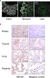
SSC4D distribution in human organs. (A) Detection of SSC4D by immunofluorescence (IF) in sections of the colon, stomach, and liver from normal human subjects. SSC4D labeling in mucous goblet cells (G) in the colon, simple columnar epithelium cells (E) in the stomach, and hepatocytes (H) in hepatic lobules is shown by arrows. Cell nuclei were stained with 4′,6-diamidino-2-phenylindole (DAPI; white). No unspecific staining was seen following incubation with secondary antibody alone, confirming specificity of the primary antibody. Scale bar, 50 μm. (B) Immunohistochemical analysis of SSC4D expression in sections of the kidney, thyroid, lung, and placenta. On the left column, SSC4D labeling was visualized by horseradish peroxidase (HRP) and substrate chromogen 3, 3'-diaminobenzidine (DAB). Positive staining of tubular epithelial cells (E) in the kidney, follicular (F) and parafollicular cells (PF) in the thyroid, pneumocytes (P) of the alveolar ducts, and of syncytiotrophoblasts (S) in the placenta is indicated by arrows. On the right column, images of sections labeled with unspecific mouse IgG mAb (negative control, sc-2025). Scale bar, 50 μm. Immunohistochemistry (IHC) and IF experiments were performed multiple times using samples from at least three different individuals.
Figure 3
Figure 3
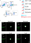
SSC4D expression in human leukocytes. (A) Flow cytometry gating strategy for the identification of monocytes, neutrophils, B cells, and CD4+ and CD8+ T cells from human blood. (B) Intracellular labeling of SSC4D in different human cell populations, visualized by flow cytometry. In the control samples, the anti-SSC4D antibody was omitted. Representative results shown are from one of four independent experiments. (C) Representative single-cell images of FACS-sorted leukocytes, immunostained for SSC4D (green) and visualized by immunofluorescence (IF). White indicates DAPI staining. Representative results shown are from one of three independent experiments using different donors.
Figure 4
Figure 4
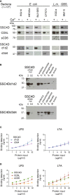
SSC4D physically binds to bacteria and bacterial endotoxins. (A) Two micrograms of each recombinant protein SSC4D, CD5L, sCD6, SSC4D-d1d2, and SSC4D-d3d4 were incubated with suspensions of 1 × 10⁸ CFU of live Escherichia coli strains BL21(DE3), RS218, IHE3034, or CFT073, Listeria monocytogenes strain EGD-e, or GBS strain BM110 in the presence or absence of Ca2+. Cell-bound proteins were detected by immunoblotting using anti-HIS mAb. Blots are representative of at least three independent experiments. (B) Recombinant SSC4D-d1d2 and SSC4D-d3d4 (2 μg each sample) were incubated with suspensions of 1 × 10⁸ CFU of live Salmonella enterica, Klebsiella pneumoniae, Enterococcus faecalis, Pseudomonas aeruginosa, or Mycobacterium avium in the presence of Ca2+. Bacteria-bound proteins were detected by immunoblotting using an anti-HIS mAb. The sensitivity of detection of this mAb for each of the recombinant hemi-forms of SSC4D can be evaluated by the detection, shown on the left side of the membranes, of 2 and 0.2 ng of purified SSC4D-d1d2 (upper blot) or 4 and 0.4 ng of purified SSC4D-d3d4 (lower blot). (C) Binding of SSC4D-d1d2 and SSC4D-d3d4 to plate-bound lipopolysaccharide (LPS) and lipoteichoic acid (LTA). Proteins were added at the indicated concentrations, and signals were detected by anti-HIS mAb followed by horseradish peroxidase (HRP)-conjugated antibody and o-Phenylenediamine dihydrochloride (OPD) substrate. Absorbance was read at 490 nm. Binding values shown were interpolated from standard curves of detection of plate-bound SSC4D-d1d2 and SSC4D-d3d4, shown in Supplementary Figure S3 . Graphs show the mean ± SD of two independent experiments performed in duplicate. (D) Binding of SSC4D, CD5L, and sCD6 to plate-bound purified LPS and LTA. Detection and measurement of binding were as in panel (C).
Figure 5
Figure 5
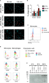
SSC4D promotes phagocytosis without binding to a ligand on human monocytes and does not induce macrophage polarization. (A) Escherichia coli pHrodo BioParticles (40 μg/ml) were added to isolated human monocytes, together with 5 μg/ml of SSC4D (middle panels) or CD5L (bottom panels), or no protein (top panels). Images were acquired for each well at 45 and 120 min after the addition of E. coli BioParticles using an IN Cell Analyzer, followed by analysis using FIJI software. Blue indicates DAPI staining, and red indicates phagocytosed E. coli BioParticles. (B) The percentage of monocytes containing E. coli BioParticles was quantified. Graph shows the mean ± SD of three independent experiments performed in duplicate. Statistical analysis was performed using Student’s t test. *p < 0.05. (C) Ex vivo monocytes were incubated with 3 μg of SSC4D or sCD6 or left unstained. Cell-bound proteins were detected with anti-HIS antibody followed with Alexa Fluor 647-conjugated anti-mouse antibody and analyzed by flow cytometry. Gray histograms represent control cells, not stained with scavenger receptor cysteine-rich (SRCR) protein but incubated with secondary antibody, red histograms represent labeling with SSC4D, and blue histograms represent labeling with sCD6. (D) Flow cytometry analysis of ex vivo monocytes (left column) and macrophage colony-stimulating factor (M-CSF)-differentiated macrophages (right column). Monocytes received for 72 h the appropriate stimuli to polarize toward M1 [interferon (IFN)-γ/lipopolysaccharide (LPS)], M2a [interleukin (IL)-4], or M2C (IL-10) subtypes. Macrophages received the same treatment, but for 24 h. CD80, CD206, and CD163 labeling confirms the polarization of monocytes and macrophages into the correct subtype. Stimulations with SSC4D or CD5L had no effect on cell polarization except for a slight effect of CD5L in polarizing macrophages into an M2a-like phenotype. Representative histograms are from one of three independent experiments. (E) SSC4D secretion upon culture infection with live bacteria. Caco-2 cells were engineered to express SSC4D fused to citrine and were cultured for 3 days at 3 × 10⁵ cells/well in a 12-well plate. Live E. coli RS218 or L. monocytogenes EGD-e were added at 1:50 multiplicity of infection (MOI). Supernatants were collected at the indicated time points, and the presence of HA-tagged SSC4D-citrine was detected by western blotting. The blot shown is representative of two independent experiments.
Figure 6
Figure 6
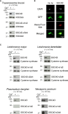
SSC4D binds to protozoan parasites. (A) Two micrograms of recombinant SSC4D, or of the hemi-forms SSC4D-d1d2 and SSC4Dd3d4, were incubated with suspensions of 1 × 10⁷ live Trypanosoma brucei brucei bloodstream forms in the presence or absence of Ca2+. Parasite-bound proteins were detected by immunoblotting using anti-HIS mAb. Membranes were reprobed with an anti-aldolase immune serum for loading control. Results shown are of one of three independent experiments. (B) Representative images of SSC4D interacting with green fluorescent protein (GFP)-expressing T. brucei. In both panels, GFP+ parasites (green) were allowed to interact with SSC4D (red), being the primary antibody omitted in the left panel, as control. DAPI (white) indicates DNA staining. The results shown are representative of four independent experiments. (C) Two micrograms of recombinant SSC4D, SSC4D-d1d2, and SSC4D-d3d4 were incubated with suspensions of 1 × 10⁷ live Leishmania major and Leishmania tarentolae promastigotes, Plasmodium berghei merozoites, and Neospora caninum tachyzoites. Interactions were detected as in panel (A), and membranes were reprobed with an anti-L. infantum cysteine synthase immune serum for loading control of L. major and L. tarentolae.

Similar articles

Cited by

References

    1. Beutler B, Jiang Z, Georgel P, Crozat K, Croker B, Rutschmann S, et al. . Genetic Analysis of Host Resistance: Toll-Like Receptor Signaling and Immunity at Large. Annu Rev Immunol (2006) 24:353–89. doi: 10.1146/annurev.immunol.24.021605.090552 - DOI - PubMed
    1. Hoving JC, Wilson GJ, Brown GD. Signalling C-Type Lectin Receptors, Microbial Recognition and Immunity. Cell Microbiol (2014) 16:185–94. doi: 10.1111/cmi.12249 - DOI - PMC - PubMed
    1. Rehwinkel J, Gack MU. RIG-I-Like Receptors: Their Regulation and Roles in RNA Sensing. Nat Rev Immunol (2020) 20:537–51. doi: 10.1038/s41577-020-0288-3 - DOI - PMC - PubMed
    1. Franchi L, Warner N, Viani K, Nunez G. Function of Nod-Like Receptors in Microbial Recognition and Host Defense. Immunol Rev (2009) 227:106–28. doi: 10.1111/j.1600-065X.2008.00734.x - DOI - PMC - PubMed
    1. Hampton RY, Golenbock DT, Penman M, Krieger M, Raetz CR. Recognition and Plasma Clearance of Endotoxin by Scavenger Receptors. Nature (1991) 352:342–4. doi: 10.1038/352342a0 - DOI - PubMed

Publication types

MeSH terms

Substances