. 2021 Dec 22:12:762032.
doi: 10.3389/fimmu.2021.762032. eCollection 2021.

Intrinsic Strand-Incision Activity of Human UNG: Implications for Nick Generation in Immunoglobulin Gene Diversification

Affiliations

Intrinsic Strand-Incision Activity of Human UNG: Implications for Nick Generation in Immunoglobulin Gene Diversification

Marina Alexeeva et al. Front Immunol. .

Abstract

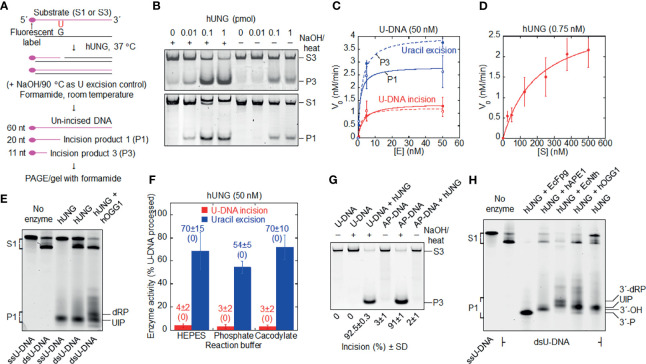
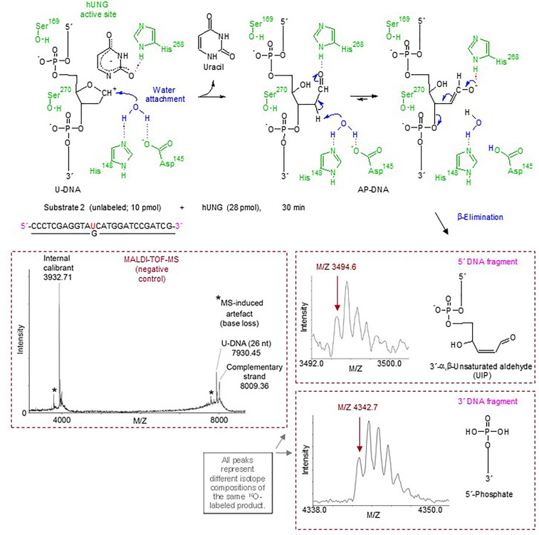
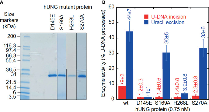
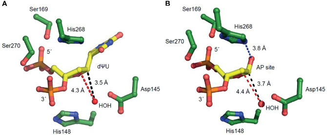
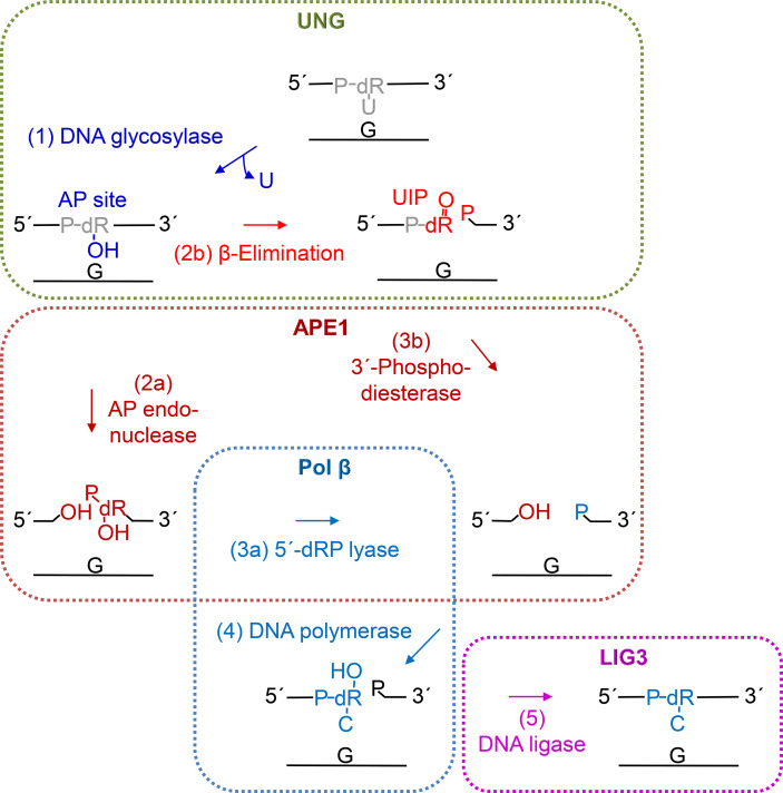
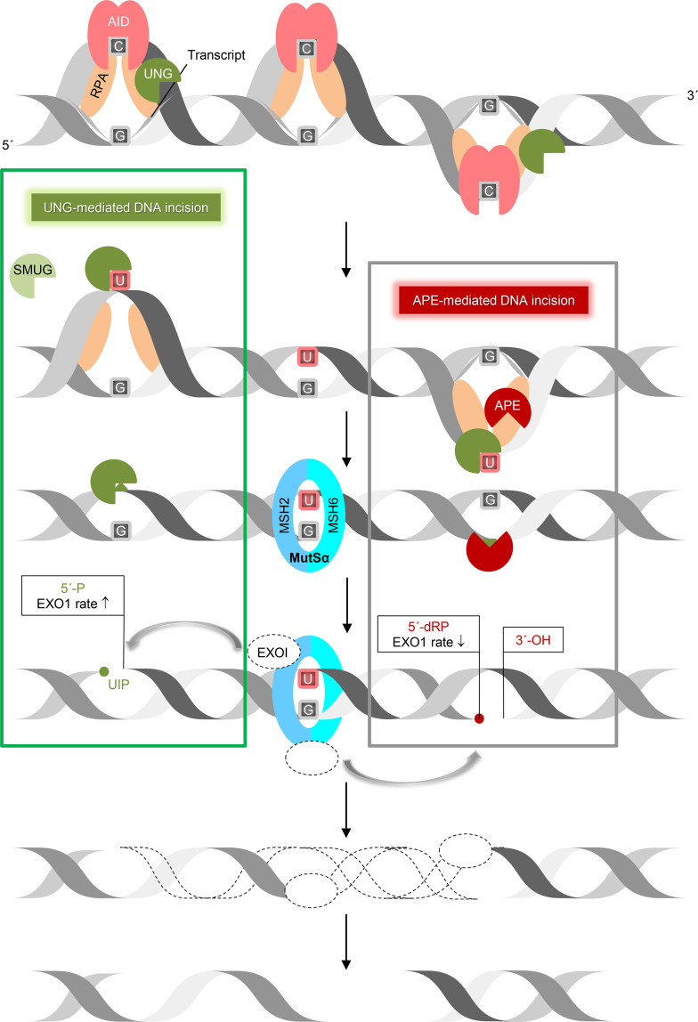
Uracil arises in cellular DNA by cytosine (C) deamination and erroneous replicative incorporation of deoxyuridine monophosphate opposite adenine. The former generates C → thymine transition mutations if uracil is not removed by uracil-DNA glycosylase (UDG) and replaced by C by the base excision repair (BER) pathway. The primary human UDG is hUNG. During immunoglobulin gene diversification in activated B cells, targeted cytosine deamination by activation-induced cytidine deaminase followed by uracil excision by hUNG is important for class switch recombination (CSR) and somatic hypermutation by providing the substrate for DNA double-strand breaks and mutagenesis, respectively. However, considerable uncertainty remains regarding the mechanisms leading to DNA incision following uracil excision: based on the general BER scheme, apurinic/apyrimidinic (AP) endonuclease (APE1 and/or APE2) is believed to generate the strand break by incising the AP site generated by hUNG. We report here that hUNG may incise the DNA backbone subsequent to uracil excision resulting in a 3´-α,β-unsaturated aldehyde designated uracil-DNA incision product (UIP), and a 5´-phosphate. The formation of UIP accords with an elimination (E2) reaction where deprotonation of C2´ occurs via the formation of a C1´ enolate intermediate. UIP is removed from the 3´-end by hAPE1. This shows that the first two steps in uracil BER can be performed by hUNG, which might explain the significant residual CSR activity in cells deficient in APE1 and APE2.

Keywords: class switch recombination; cytosine deamination; human UNG; immunoglobulin diversification; somatic hypermutation.


Conflict of interest statement

The authors declare that the research was conducted in the absence of any commercial or financial relationships that could be construed as a potential conflict of interest.

Figures

Figure 1
Figure 1
Formation of uracil-DNA incision product (UIP) by hUNG. (A) DNA substrate and base excision assay. Either substrate 1 (S1) or substrate 3 (S3; the base sequences of their labeled strands are presented in Materials and Methods) was exposed to hUNG, resulting in incision product 1 (P1) or incision product 3 (P3), respectively. hUNG (1 pmol) was incubated 10 min with DNA substrate (1 pmol) at 37°C in 45 mM HEPES [4-(2-hydroxyethyl)-1-piperazineethane-sulfonic acid]–KOH, pH 7.8, 0.4 mM EDTA, 1 mM DTT, 70 mM KCl, and 2% (v/v) glycerol (reaction buffer; final volume, 20 µl), if not otherwise stated. (B) Protein dependence of dsU-DNA incision (red) and uracil excision (blue). PAGE was performed at 100 V for 30 min (upper panel) or 50 min (lower panel) using a 12% (w/v) gel, which contained 3% (v/v) formamide, where (C) presents the three to four independent measurements performed. (D) Incision of dsU-DNA by hUNG follows Michaelis Menten kinetics. hUNG (0.015 pmol) was incubated with an increasing concentration of S1 for 20 min, where the other experimental details are the same as in (B) [except that a 20% (w/v) gel was used]; each value represents the average of 7 independent measurements. (E) Cleavage of ss- and dsU-DNA by hUNG. S1 or its labeled strand was incubated with hUNG alone for 20 min (third and fourth lane), which in one case was followed by incubation together with hOGG1 (3 pmol) for additional 30 min (fifth lane). PAGE was performed at 300 V for 7 h using a 20% (w/v) gel containing 7 M urea. (F) Incision of dsU-DNA by hUNG in different buffers. S1 was incubated with and without hUNG in reaction buffer (HEPES), 45 mM sodium cacodylate, or 45 mM potassium phosphate, with the same pH and additions as for reaction buffer (see Supplementary Figure 1 for details). In each case, five independent measurements were performed. Each value represents the average ± SD, where control value without enzyme is shown in parenthesis. (G) No cleavage of dsAP-DNA (U replaced by AP site) by hUNG. dsU-DNA (S3, 1 pmol) was converted to dsAP-DNA by incubation with EcUng (2 pmol) at 37°C for 10 min. AP- or U-DNA was incubated with and without hUNG. PAGE was performed at 100 V for 1 h using a 12% (w/v) gel, which contained 3% (v/v) formamide. The inability of the NaOH/heat treatment to cleave U-DNA as opposed to AP-DNA verified the integrity of the former as well as the nature of the latter. The complete cleavage of U-DNA by the NaOH/heat treatment following hUNG exposure verified active enzyme. It should be noted that we always detected UIP in our samples of AP-DNA, which was difficult to avoid since we routinely used EcUng for its preparation. In the AP-DNA sample presented in the figure, we succeeded to wash away most UIP during the two precipitation steps employed. Three independent experiments with virtually the same result were performed. (H) Indirect identification of UIP by its electrophoretic mobility compared to that of characterized enzymes. hUNG (1 pmol) was incubated with S1 (1 pmol); either alone for 20 min (seventh lane), together with EcFpg (4 pmol) for 30 min (third lane), alone for 20 min and thereafter together with hAPE1 (0.45 pmol) for an additional 30 min (fourth lane), together with EcNth (1 pmol) for 30 min (fifth lane), or alone for 20 min and thereafter together with hOGG1 (4 pmol) for an additional 30 min (sixth lane). Different incision products were separated from un-incised DNA by PAGE at 300 V for 7 h using a 20% (w/v) gel containing 7 M urea. The experiment with the arrangement presented was performed three times. More than 10 additional experiments were performed with other arrangements to indicate the 3´ incision product. dRP, deoxyribose phosphate; nt, nucleotides; P, phosphate.
Figure 2
Figure 2
Suggested hUNG reaction mechanism for incision of U-DNA into UIP and 5´-phosphate as identified by MALDI-TOF-MS and supported by structural modeling and site-directed mutagenesis. In the upper panel, amino acid residues of hUNG involved in catalysis are indicated in green; their hydrogen bonds with catalytic water and substrate are indicated by red dotted lines. Enzymatically activated H2O (in blue) is attached at C1´ of the abasic deoxyribose [blue arrow; based on refs. (49, 50)]. Proposed electronic (that cause atomic) transfers that are involved in the formation of UIP are indicated by short blue arrows. A hydrogen bond between His268 and the formyl oxygen of the ring-opened abasic deoxyribose (51) is proposed. We suggest that Asp145 acts as a general base, and that the attraction of electrons by the C1´ formyl group improves the C2´ hydrogen as a leaving group. The middle and lower right panels show the MALDI-TOF-MS signals of the 5´ and 3´ DNA incision fragments, respectively, while the middle left panel shows the signals obtained from incubation without enzyme.
Figure 3
Figure 3
U-DNA incision and uracil excision activity of hUNG mutant proteins purified to apparent physical homogeneity. (A) SDS-PAGE of purified proteins stained with Coomassie Blue. Samples (20 µl; treated 5 min at 95°C in NuPAGE® LDS Sample Buffer, Cat. No. NP0007, Life Technologies) and protein markers (Mark12™ Unstained Standard, Cat. No. LC5677, Life Technologies) were run on a 4–20% Mini-PROTEAN® TGX™ Precast Gel (12 wells; Bio-Rad) in 25 mM Tris, 0.192 M glycine, 0.1% (w/v) SDS at 200 V for 35 min. Left lane, protein markers; right lanes, hUNG mutant proteins. (B) Different decrease in U-DNA incision and uracil excision activity by site-directed mutant hUNG proteins. Wild-type and mutant proteins (0.015 pmol) were incubated with S1 (10 pmol) at 37°C for 20 min (see Figure 1A). Each value represents the average ± SD of 5–16 independent measurements.
Figure 4
Figure 4
Amino acid residues of hUNG active site region positioned to participate in uracil excision and uracil-DNA incision. These enlarged views in which 2´-deoxyuridine is replaced with (A) 2´-deoxypseudouridine (dΨU) or (B) an AP site indicate amino acid residues involved in substrate binding and coordination. The distances between the catalytic water molecule and the site C1´ where it is attached (causing uracil excision) and the site C2´ of proton elimination (causing uracil-DNA incision) are indicated by black and red broken lines, respectively. The His268 NH–C1´O distance in (B), facilitating proton removal from C2´, is indicated by a blue broken line.
Figure 5
Figure 5
Proposed steps of hUNG-initiated uracil BER. After uracil is removed (step 1, blue) by the DNA glycosylase activity of UNG (green), the AP site is mostly incised (step 2a) by APE1 (dark red) leaving behind a 3´-OH group. Then, the 5´-deoxyribose (dR) phosphate (P) remnant is removed by the dRP lyase activity (step 3a) of DNA polymerase β (Pol β, light blue), following conclusion of BER by insertion of the correct dCMP (step 4) by Pol β and nick-sealing (step 5) by DNA ligase III (LIG3, purple). Alternatively, UNG may itself incise the AP site (step 2b, red) by β-elimination (Figure 2, upper middle and right panels) leaving behind a 3´-α,β-unsaturated aldehyde (UIP; Figure 2, middle right panel), which can be removed by the 3´-phosphodiesterase activity (step 3b) of APE1, and a 5´-P (Figure 2, lower right panel).
Figure 6
Figure 6
UNG-mediated DNA incision in CSR. This working model suggests how removal of AID-generated uracil followed by incision of the AP site by UNG and nick processing by exonuclease 1 (EXO1) form DSB in immunoglobulin switch regions. Transcription of the targeted immunoglobulin gene region forms bubbles in DNA, so granting AID access to ssDNA (stabilized by RPA) to deaminate C to U. This results in UNG recruitment (by RPA) and uracil excision. According to our results (left square), UNG (with SMUG1 as a backup) is able to incise the AP site, leaving behind a 5´-phosphate, which is a better substrate for exonuclease 1 (EXO1) 5´→ 3´ digestion than the 5´-deoxyribose phosphate left behind by APE1 incision (right square). This model relies on the MMR component MutSα (MSH2/6), which recognizes a U:G mismatch and recruits EXO1. This also applies to ssDNA patch generation by EXO1 in SHM. ↑, increased, ↓, decreased.

Similar articles

Cited by

References

    1. Friedberg EC, Walker GC, Siede W, Wood RD, Schultz RA, Ellenberger T. DNA Repair and Mutagenesis. 2nd ed. Washington, DC: ASM Press; (2006).
    1. Lindahl T. Instability and Decay of the Primary Structure of DNA. Nature (1993) 362:709–15. doi: 10.1038/362709a0 - DOI - PubMed
    1. Lucas-Lledó JI, Maddamsetti R, Lynch M. Phylogenomic Analysis of the Uracil-DNA Glycosylase Superfamily. Mol Biol Evol (2011) 28:1307–17. doi: 10.1093/molbev/msq318 - DOI - PMC - PubMed
    1. Bauer NC, Corbett AH, Doetsch PW. The Current State of Eukaryotic DNA Base Damage and Repair. Nucleic Acids Res (2015) 43:10083–101. doi: 10.1093/nar/gkv1136 - DOI - PMC - PubMed
    1. Kim Y-J, Wilson DM, 3rd. Overview of Base Excision Repair Biochemistry. Curr Mol Pharmacol (2012) 5:3–13. doi: 10.2174/1874467211205010003 - DOI - PMC - PubMed

Publication types