. 2021 Dec 23:12:781857.
doi: 10.3389/fpls.2021.781857. eCollection 2021.

Chloroplast Ribosomes Interact With the Insertase Alb3 in the Thylakoid Membrane

Affiliations

Chloroplast Ribosomes Interact With the Insertase Alb3 in the Thylakoid Membrane

Bernd Ackermann et al. Front Plant Sci. .

Abstract

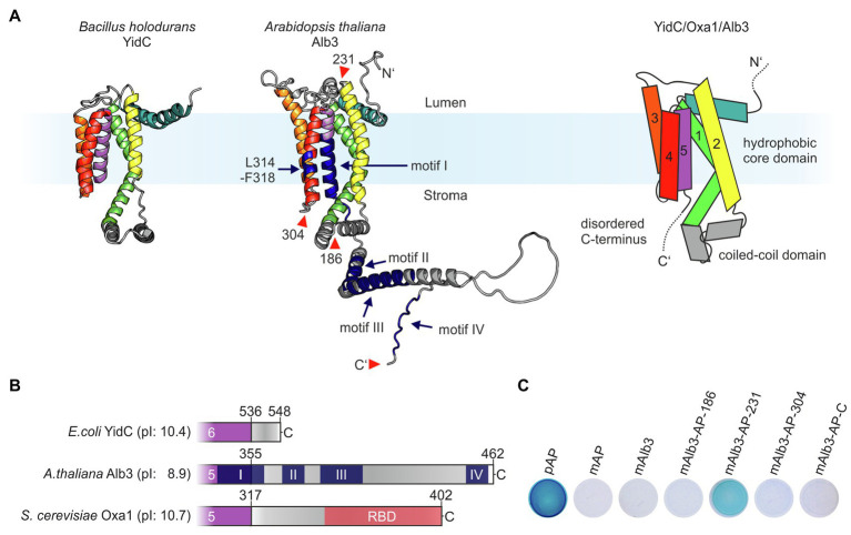
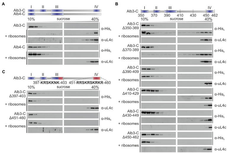
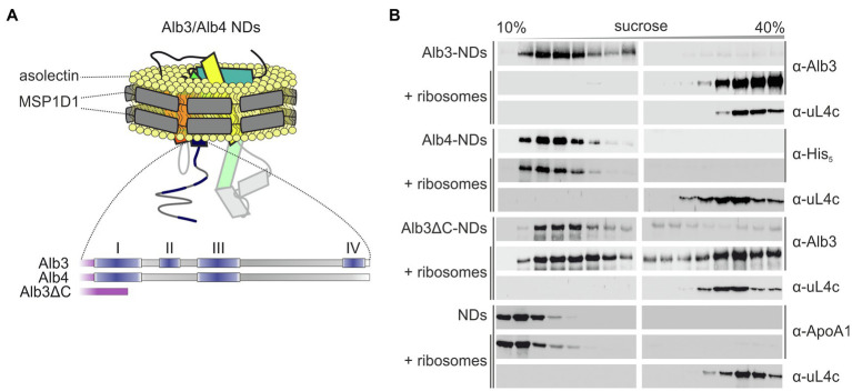
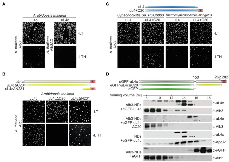
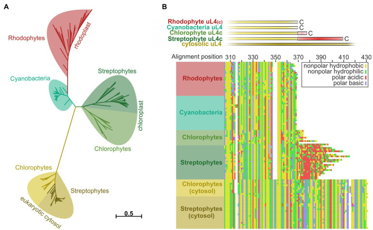
Members of the Oxa1/YidC/Alb3 protein family are involved in the insertion, folding, and assembly of membrane proteins in mitochondria, bacteria, and chloroplasts. The thylakoid membrane protein Alb3 mediates the chloroplast signal recognition particle (cpSRP)-dependent posttranslational insertion of nuclear-encoded light harvesting chlorophyll a/b-binding proteins and participates in the biogenesis of plastid-encoded subunits of the photosynthetic complexes. These subunits are cotranslationally inserted into the thylakoid membrane, yet very little is known about the molecular mechanisms underlying docking of the ribosome-nascent chain complexes to the chloroplast SecY/Alb3 insertion machinery. Here, we show that nanodisc-embedded Alb3 interacts with ribosomes, while the homolog Alb4, also located in the thylakoid membrane, shows no ribosome binding. Alb3 contacts the ribosome with its C-terminal region and at least one additional binding site within its hydrophobic core region. Within the C-terminal region, two conserved motifs (motifs III and IV) are cooperatively required to enable the ribosome contact. Furthermore, our data suggest that the negatively charged C-terminus of the ribosomal subunit uL4c is involved in Alb3 binding. Phylogenetic analyses of uL4 demonstrate that this region newly evolved in the green lineage during the transition from aquatic to terrestrial life.

Keywords: Oxa1/YidC/Alb3 protein family; cotranslational protein transport; nanodiscs; ribosomes; thylakoid membrane biogenesis.


Conflict of interest statement

The authors declare that the research was conducted in the absence of any commercial or financial relationships that could be construed as a potential conflict of interest.

Figures

Figure 1
Figure 1
Proteins of the Oxa1 family share common structural features. (A) Comparison of the crystal structure of YidC from Bacillus halodurans (left; PDB: 3WO6; Kumazaki et al., 2014a) and the AlphaFold model of Arabidopsis thaliana Alb3 without signal peptide (middle; model: AF-Q8LBP4-F1; Jumper et al., 2021). Regions in the structural model of Alb3 highlighted in dark blue indicate conserved sequence motifs involved in cpSRP43 binding (residues 314–318 within the fourth TMH and motifs II and IV in the C-terminal region); the conserved motifs I and III are also highlighted in dark blue. The red arrowheads indicate the alkaline phosphatase (AP) insertion sites mentioned in C. Right: scheme of the predicted topology of the hydrophobic core region of members of the Oxa1 family encompassing five transmembrane helices. (B) Features of the C-terminal sequences of members of the Oxa1 family. The start of the C-terminal regions were defined to the first residue after the last transmembrane helix according to the crystal structure of Escherichia coli YidC (PDB: 3WVF; Kumazaki et al., 2014b) and the Alphafold models of A. thaliana Alb3 (see above) and Saccharomyces cerevisiae Oxa1 (model: AF-P39952-F1; Jumper et al., 2021). Theoretical pI-values for the indicated C-terminal peptide sequences were calculated using ExPASy ProtParam. Prominent sequence motifs are marked. I, II, III, and IV: conserved motifs of the Alb3 C-terminal region (Falk et al., 2010); RBD: conserved helical ribosome binding domain of the Oxa1 C-terminal region (Jia et al., 2003). (C) Alkaline phosphatase (AP) activity of bacterial cells carrying expression plasmids coding for different Alb3-AP fusions to identify membrane protein topology. AP was fused into mature Alb3 at amino acid positions 186 (mAlb3-AP-186), 231 (mAlb3-AP-231), 304 (mAlb3-AP-304), and to the C-terminus (mAlb3-AP-C). Control cells expressed the precursor of AP (pAP), mature AP (mAP) and mAlb3. The positions of the fusions are indicated schematically in A (red arrowheads).
Figure 2
Figure 2
Sucrose gradient interaction assay of recombinant Alb3 and Alb4 C-terminus variants with chloroplast ribosomes. The recombinant His-tagged C-terminal regions of Alb3 and Alb4 and the indicated deletion constructs were incubated with chloroplast ribosomes and loaded onto a sucrose density gradient. After ultracentrifugation, the gradient fractions were analyzed immunologically using antibodies against the His-tag (α-His) and the chloroplast ribosomal protein uL4c (α-uL4c). Sucrose density gradient centrifugation of the recombinant proteins in the absence of ribosomes was used as negative control. Alb3-C, C-terminal region of Alb3 (amino acids 350–462); Alb4-C, C-terminal region of Alb4 (amino acids 334–499). (A) Comparison of Alb3-C and Alb4-C. (B) A successive experimental series of 20 amino acid deletions introduced into the Alb3-C construct. (C) Targeted deletions of highly conserved arginine- and lysine-rich sequences within motif III and motif IV.
Figure 3
Figure 3
Sucrose gradient interaction assay of Alb3 variants and Alb4 reconstituted in nanodiscs with chloroplast ribosomes. (A) Schematic representation of recombinant His-tagged full-length mature Alb3 (amino acids 55–462), Alb3ΔC (amino acids 55–369) and Alb4 (amino acids 46–499) reconstituted in MSP1D1 nanodiscs (NDs; not to scale) with asolectin as bilayer forming lipid. (B) Nanodiscs (NDs) containing Alb3 (Alb3-NDs), Alb4 (Alb4-NDs), or Alb3ΔC (Alb3ΔC-NDs) and NDs without inserted membrane protein as negative control were incubated with chloroplast ribosomes and loaded onto a sucrose density gradient. After ultracentrifugation, the gradient fractions were analyzed immunologically using antibodies against the His-tag (α-His), A. thaliana chloroplast Alb3 (α-Alb3), human Apolipoprotein A1 (α-ApoA1; the wildtype template of MSP1D1) and the chloroplast ribosomal protein uL4c (α-uL4c). Sucrose density gradient centrifugation of the Alb constructs, containing NDs in the absence of ribosomes was used as negative control.
Figure 4
Figure 4
Split ubiquitin and size exclusion chromatography interaction assays between Alb3 and uL4c constructs. Split ubiquitin assays (A–C) were performed employing fusions to the C- (Cub) and N- (NubG) terminal halves of ubiquitin. The Alb3 constructs were C-terminally fused to Cub and the uL4 constructs were N-terminally fused to NubG. Alg5-NubI and Alg5-NubG served as positive and negative controls, respectively. Yeast colonies were plated on permissive medium (-LT) and on selective medium (-LTH). A full version with all control experiments is shown in Supplementary Figure S2. (A) Comparison between the interaction of mature Alb3 (amino acids 55–462) and C-terminally truncated Alb3 (amino acids 55–369; Alb3ΔC) with A. thaliana uL4c. (B) Interaction assay between Alb3 and uL4c or uL4c deletion constructs lacking 20 C-terminal amino acids (uL4cΔC20) or 231 N-terminal amino acids resulting in a construct coding for the last 50 amino acids (uL4cΔN231). (C) Interaction assay between Alb3 and cyanobacterial uL4 proteins or fusions of cyanobacterial uL4 proteins with the C-terminal 20 amino acids of A. thaliana uL4c (uL4+C20). (D) Size exclusion chromatography assays were performed by incubating Alb3-NDs with fusion constructs of eGFP and the C-terminal half of A. thaliana uL4c with and without its negative C-terminus, eGFP-uL4c (amino acids 150–282) and eGFP-uL4cΔC20 (amino acids150–262), respectively. Control assays were conducted using Alb3-NDs in combination with eGFP and empty NDs in combination with eGFP-uL4c. The elution fractions between 7.5 and 18.5 ml were analyzed immunologically using antibodies against A. thaliana chloroplast Alb3 (α-Alb3), A. thaliana chloroplast ribosomal protein uL4c (α-uL4c), Human Apolipoprotein A1 (α-ApoA1; the wildtype template of MSP1D1) and eGFP (α-GFP). Asterisks mark C-terminal degradation bands of the eGFP-uL4c-constructs.
Figure 5
Figure 5
Reconstruction of phylogenetic relationships and amino acid sequence comparisons between uL4 proteins of photosynthetic organisms. Phylogeny- and multiple sequence alignment-based analysis of plastidic and cytosolic uL4 sequences from chlorophytes, streptophytes, rhodophytes, and cyanobacteria. (A) Unrooted maximum likelihood tree of the homologous uL4 peptide sequence regions. Branch lengths are scaled according to the number of substitutions per site. (B) MAFFT alignment of the uL4 amino acid sequences, using the L-INS-I algorithm. Top: schematic representation of the aligned sequences. Bottom: Cropped alignment emphasizing the C-terminal regions of the bacterial/plastidic uL4/uL4c proteins. Amino acids are represented by their chemical properties: non-polar hydrophobic (yellow; A,F,I,L,M,P,V,W), non-polar hydrophilic (green; C,G,N,Q,S,T,Y), polar basic (blue; H,K,R) and polar acidic (red; D,E).

References

    1. Bals T., Dünschede B., Funke S., Schünemann D. (2010). Interplay between the cpSRP pathway components, the substrate LHCP and the translocase Alb3: an in vivo and in vitro study. FEBS Lett. 584, 4138–4144. doi: 10.1016/j.febslet.2010.08.053, PMID: - DOI - PubMed
    1. Bedard J., Trosch R., Wu F., Ling Q., Flores-Perez U., Topel M., et al. (2017). Suppressors of the chloroplast protein import mutant tic40 reveal a genetic link between protein import and thylakoid biogenesis. Plant Cell 29, 1726–1747. doi: 10.1105/tpc.16.00962, PMID: - DOI - PMC - PubMed
    1. Benz M., Bals T., Gugel I. L., Piotrowski M., Kuhn A., Schünemann D., et al. (2009). Alb4 of Arabidopsis promotes assembly and stabilization of a non chlorophyll-binding photosynthetic complex, the CF1CF0-ATP synthase. Mol. Plant 2, 1410–1424. doi: 10.1093/mp/ssp095, PMID: - DOI - PubMed
    1. Chandrasekar S., Shan S. O. (2017). Anionic phospholipids and the albino3 translocase activate signal recognition particle-receptor interaction during light-harvesting chlorophyll a/b-binding protein targeting. J. Biol. Chem. 292, 397–406. doi: 10.1074/jbc.M116.752956, PMID: - DOI - PMC - PubMed
    1. Denisov I. G., Grinkova Y. V., Lazarides A. A., Sligar S. G. (2004). Directed self-assembly of monodisperse phospholipid bilayer nanodiscs with controlled size. J. Am. Chem. Soc. 126, 3477–3487. doi: 10.1021/ja0393574, PMID: - DOI - PubMed

LinkOut - more resources