. 2021 Aug 26:60:e47.
doi: 10.6620/ZS.2021.60-47. eCollection 2021.

Classification and Morphology of Rhinocypha spp. (Odonata): A Comprehensive Taxonomic Study Within the Females

Affiliations

Classification and Morphology of Rhinocypha spp. (Odonata): A Comprehensive Taxonomic Study Within the Females

Noorhidayah Mamat et al. Zool Stud. .

Abstract

Studies on Odonata have gained attention worldwide as well as locally in Malaysia. Although there is a wealth of data available to be utilized for solving taxonomic problems, ecological and behavioural research areas are more favoured than taxonomy and systematics. Thus, there are confusions over how to correctly identify closely related and sympatric species, especially in female odonates. One such example is in the genus Rhinocypha. Consequently, the present study focuses on taxonomic work, employing multi-approaches in the form of morphological (morphological diagnostics, Field Emission Scanning Electron Microscope (FESEM) and geometric morphometric analysis), applying the molecular technique. Seventeen morphological characteristics were created to differentiate between the females of Rhinocypha spp. A FESEM was used on the female's ovipositor to focus on the anal appendages and sheathing valve (V3). Also, the phylogenetic patterns expressed by COI and 16S rRNA genes, and canonical variate analysis for the wing geometric morphometric revealed three clusters that supported the distinction of the Rhinocypha group. In summary, this study effectively developed an integrated approach of classic morphological and trendy molecular, combined with FESEM microscopy techniques, which provided corroborative evidence and resolved taxonomic uncertainties.

Keywords: 16S rRNA; Dragonflies; Female’s ovipositor; Geometric morphometric; Mitochondrial COI.


Figures

Fig. 1.
Fig. 1.
Lateral view of thorax and anterior abdomen. Characters used the morphological nomenclature by Djikstra et al. (2014) and modified in order to create the key identification for females of Rhinocypha spp. using from Gunther (2009).
Fig. 2.
Fig. 2.
Lateral view of the external morphology of the ovipositor of Rhinocypha spp. Ap: anal appendages; St: stylus; Dt: distal tooth; V3: third valves of ovipositor (valvulae 3); Lam: basal plate of ovipositor (lamina valvarum).
Fig. 3.
Fig. 3.
Landmark configuration of Rhinocypha spp. Fifteen landmarks were used in geometric morphometric analysis. Landmarks represent: (1) costa –subcostal connection, (2, 3 & 4) distal angles of arculus, (5) the nodus, (6) posterior intersection of the pterostigma and radius 1 (R1), (7) end of vein radius 2 (R2), (8) posterior end of the radius 4 (R4), (9) posterior end of the anterior media (MA), (10) posterior end of the Cubital Vein (CuP), (11) posterior end of the Anal Vein 1 (A1), (12) proximal apex of anal triangle, (13) anterior end of the cubital vein supplementary (Cupspl); (14) anterior end of the anterior media supplementary (Mspl); and (15) anterior end of the radius 4 supplementary (R4spl).
Fig. 4.
Fig. 4.
Wing of Rhinocypha spp. (a) Rhinocypha fenestrella, (b) Rhinocypha perforata, and (c) Rhinocypha biforata.
Fig. 5.
Fig. 5.
Thorax of the females of Rhinocypha spp. (a) R. fenestrella, (b) R. perforata, and (c) R. biforata.
Fig. 6.
Fig. 6.
Scanning electron micrographs of the morphometric measurements of the ovipositor for the females of Rhinocypha spp. (lateral view). (a) Rhinocypha biforata, (b) Rhinocypha fenestrella, (c) Rhinocypha perforata. (i, ii & iii) length of each segment; (iv) length of anal appendages; (v) basal width of anal appendages; (vi) length of stylus; (vii) width of the V3.
Fig. 7.
Fig. 7.
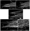
Scanning electron micrographs of anal appendages of Rhinocypha spp. (a) Rhinocypha biforata –inset indicates the group of sensilla; (b) Rhinocypha fenestrella –inset shows the caeloconica-like sensilla; (c) Rhinocypha perforata; (d) group of sensilla on the tip of anal appendages; (e) caeloconica-like sensilla on the surface of the anal appendages. Gs: group of sensilla; Bs: basiconic sensilla; As: articulated setae.
Fig. 8.
Fig. 8.
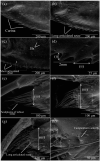
Scanning electron micrographs of sheathing valve (V3) and distal tooth of Rhinocypha spp. (a) Rhinocypha biforata, (b) Rhinocypha fenestrella, (c) Rhinocypha perforata –inset shows the carina, (d) measurement of (i) the peak of the tooth to the median base (ii) the space between the tooth. (e, f & g) shows the scanning electron micrographs of distal tooth of Rhinocypha spp.: (e) Rhinocypha biforata -(iii) indicates the width of the distal tooth, (f) Rhinocypha fenestrella –inset shows the campaniform sensilla, (g) Rhinocypha perforata, (h) campaniform sensilla at the distal tooth surface.
Fig. 9.
Fig. 9.
Scanning electron micrographs of the stylus and the base of stylus of Rhinocypha spp. (a) Rhinocypha biforata, (b) Rhinocypha fenestrella –(i) width of the stylus from the third of hair sensilla, (c) Rhinocypha perforata. (d, e & f) scanning electron micrographs of the base of stylus. (d) Rhinocypha biforata, (e) Rhinocypha fenestrella, (f) Rhinocypha perforata.
Fig. 10.
Fig. 10.
Scatterplot of all 15 landmarks configurations after Procrustes superimposition. The plotted line and blue dots represent the mean shape for the respective species; (a) Rhinocypha biforata, (b) Rhinocypha fenestrella, (c) Rhinocypha perforata.
Fig. 11.
Fig. 11.
Wireframe visualization of shape variation along the principal components one (PC1) from geometric morphometric analysis. (a) Rhinocypha biforata, (b) Rhinocypha fenestrella, (c) Rhinocypha perforata. Light blue landmarks represent the configuration of average specimen; dark blue landmarks represent one approximate extreme of the variation on that axis. Percentages indicate the proportion of total variance explained by each axis.
Fig. 12.
Fig. 12.
Results of principal components analysis of all specimens. PC1 = 48.64%, PC2 = 17.45%, accounting for 66.09% of the total variation.
Fig. 13.
Fig. 13.
Thin-plate spline deformation grids of wing shape variation in Rhinocypha spp. (a) Rhinocypha biforata, (b) Rhinocypha fenestrella, (c) Rhinocypha perforata, demonstrating the directions (arrows).
Fig. 14.
Fig. 14.
Canonical Variate analysis (CVA) plot. CV1 (eigenvalue 8.887) vs CV2 (eigenvalue 2.150). 90% confidence ellipses of CVA scores. Colour of ellipses corresponds to the species written alongside
Fig. 15.
Fig. 15.
Maximum Likelihood (ML) phylogenetic tree of Rhinocypha spp. based on (a) COI gene, (b) 16S rRNA gene, and (c) combined COI + 16S rRNA sequences with R. bisignata as an outgroup. Bootstrap values are shown on the branches.

Similar articles

Cited by

References

    1. Adams DC, Rohlf FJ, Slice D. 2004. Geometric morphometrics: Ten years of progress following the ‘revolution’. Ital J Zool 71:5–16. doi:10.1080/11250000409356545.
    1. Alexander LC, Delion M, Hawthorne DJ, Lamp WO, Funk DH. 2009. Mitochondrial lineages and DNA barcoding of closely related species in the mayfly genus Ephemerella (Ephemeroptera: Ephemerellidae). J North Am Benthol Soc 28:584–595. doi:10.1899/08-150.1.
    1. Andrew CG. 1966. Sexual recognition in adult Erythemis simplicicollis (Odonata: Anisoptera). Ohio J Sci 66:613–617.
    1. Asahina S. 1954. A morphological study of a relict dragonfly Epiophlebia superstes Selys (Odonata, Anisozygoptera). Society for the Promotion of Science. Tokyo, Japan.
    1. Balke M, Wewalka G, Alarie, Ribera I. 2007. Molecular phylogeny of Pacific Island Colymbetinae: Radiation of New Caledonian and Fijian species (Coleoptera, Dytiscidae). Zool Scr 36:173–200. doi:10.1111/j.1463-6409.2006.00265.x.

LinkOut - more resources