

Primary intraventricular gliosarcoma on MRI: A challenging diagnosis
- PMID: 35003460
- PMCID: PMC8718817
- DOI: 10.1016/j.radcr.2021.12.022
Primary intraventricular gliosarcoma on MRI: A challenging diagnosis
Abstract
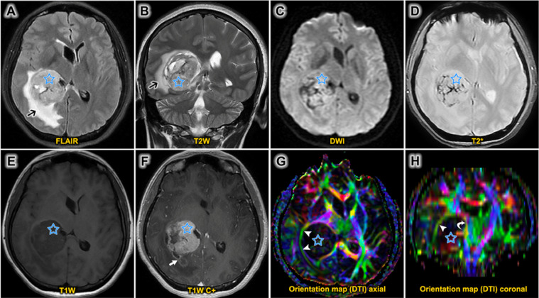
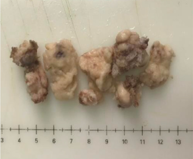
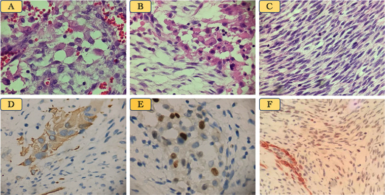
Gliosarcoma (GS) is an uncommon central nervous system tumor with several characteristics of a malignant neoplasm and poor prognosis. The majority of GS reports describe a predilection for the cerebral hemispheres, and cases of intraventricular GS are extremely rare, with only a few reported. In addition, intraventricular GS has not been associated with any unique radiographic or clinical features, which can result in misdiagnosis as other intraventricular tumor types. In this report, we present the case of a 32-year-old woman with GS in the trigone of the lateral ventricle and provide a retrospective review of similar, previously reported cases.
Keywords: Gliosarcoma; Intraventricular; Magnetic resonance imaging.
© 2021 The Authors. Published by Elsevier Inc. on behalf of University of Washington.
Figures
Similar articles
-
A Rare Tumor in a Rare Location: Radiology and Pathology Findings With a Literature Review on Intraventricular Gliosarcoma.Cureus. 2023 Feb 4;15(2):e34622. doi: 10.7759/cureus.34622. eCollection 2023 Feb. Cureus. 2023. PMID: 36891012 Free PMC article.
-
Primary gliosarcoma with long-survival: report of two cases and review of literature.Int J Clin Exp Pathol. 2014 Aug 15;7(9):6323-32. eCollection 2014. Int J Clin Exp Pathol. 2014. PMID: 25337286 Free PMC article. Review.
-
An unusual case of intraventricular gliosarcoma.Clin Neuropathol. 2009 Sep-Oct;28(5):379-83. doi: 10.5414/npp28379. Clin Neuropathol. 2009. PMID: 19788054 Review.
-
Intraventricular Gliosarcomas: Literature Review and a Case Description.World Neurosurg. 2016 Jun;90:707.e5-707.e12. doi: 10.1016/j.wneu.2016.03.033. Epub 2016 Mar 19. World Neurosurg. 2016. PMID: 27004757 Review.
-
IDH1-mutant primary intraventricular gliosarcoma: Case report and systematic review of a rare location and molecular profile.Surg Neurol Int. 2020 Nov 6;11:372. doi: 10.25259/SNI_586_2020. eCollection 2020. Surg Neurol Int. 2020. PMID: 33408906 Free PMC article.
Cited by
-
A Rare Tumor in a Rare Location: Radiology and Pathology Findings With a Literature Review on Intraventricular Gliosarcoma.Cureus. 2023 Feb 4;15(2):e34622. doi: 10.7759/cureus.34622. eCollection 2023 Feb. Cureus. 2023. PMID: 36891012 Free PMC article.
References
-
- Stroebe H. Ueber entstehung und bauder gehirnglioma. Beitr Pathol Anat. 1895;19:405–486.
-
- Louis DN, Ohgaki H, Wiestler OD, Cavenee WK, Burger PC, Jouvet A, et al. The 2007 WHO classification of tumours of the central nervous system. Acta Neuropathol. 2007;114(2):97–109. doi: 10.1007/s00401-007-0243-4. Epub 2007 Jul 6. Erratum in: Acta Neuropathol. 2007 Nov;114(5):547. PMID: 17618441; PMCID: PMC1929165. - DOI - PMC - PubMed
-
- Han SJ, Yang I, Tihan T, Prados MD, Parsa AT. Primary gliosarcoma: key clinical and pathologic distinctions from glioblastoma with implications as a unique oncologic entity. J Neurooncol. 2010;96(3):313–320. doi: 10.1007/s11060-009-9973-6. Epub 2009 Jul 18. PMID: 19618114; PMCID: PMC2808523. - DOI - PMC - PubMed
-
- de Macedo Filho LJM, Barreto EG, Martins PLB, Filho ENS, Gerson G, de Albuquerque LAF. IDH1-mutant primary intraventricular gliosarcoma: Case report and systematic review of a rare location and molecular profile. Surg Neurol Int. 2020;11:372. doi: 10.25259/SNI_586_2020. PMID: 33408906; PMCID: PMC7771479. - DOI - PMC - PubMed
Publication types
LinkOut - more resources
Full Text Sources
