

Historical Aspects of DDH
- PMID: 35003531
- PMCID: PMC8688643
- DOI: 10.1007/s43465-021-00470-x
Historical Aspects of DDH
Abstract
Background: Little was known about developmental dysplasia of the hip (DDH) in the early historical era. Symptoms such as limping were caused by a variety of disease processes, many of which were life threatening. It was not until the discovery of X-ray in 1896 that clear understanding of childhood hip conditions, including DDH, could evolve.
Methods: We reviewed available literature and distilled it into this summary of the history of our understanding of DDH.
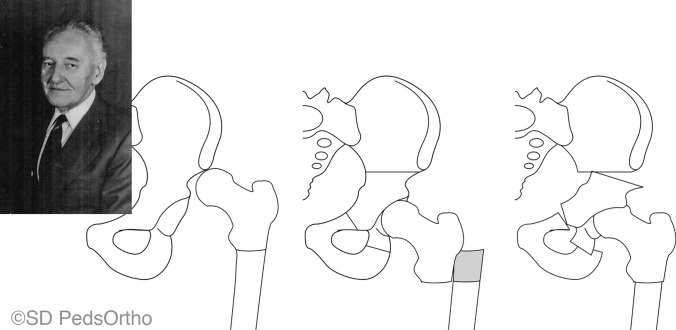
Results: The development of non-operative methods relied on plaster of Paris cast reductions and later splints and harness systems leading to the Pavlik harness (1950's). The development of ultrasound as a diagnostic technique made early diagnosis and treatment possible. Surgical approaches to DDH treatment required several key discoveries: invention of general anesthesia (1840s); development of sterilization techniques (1860-1880); discovery of X-ray (1890s); development of stainless steel (1920s); and the discovery of antibiotics (1930s). These surgical advances allowed the development of a remarkable variety of operations to treat DDH including open reduction techniques, and osteotomies of the acetabulum and proximal femur.
Conclusion: The path to accurate diagnosis and predictable treatment of DDH parallels the many advances that application of the scientific method has allowed in the specialty of orthopedic surgery. The development of academic centers that focus on research and education in childhood hip disorders, as well as a growing number of centers that focus on hip problems in adolescents and young adults, assure a continuous and changing "history" of this common childhood hip condition.
Keywords: DDH; History; Pediatric orthopedics.
© Indian Orthopaedics Association 2021.
Conflict of interest statement
Conflict of interestNo external funding was received for this paper. This paper was supported by the Rady Children’s Hospital, San Diego Division of Orthopedics. Author JDB has nothing to disclose. Author DRW has the following disclosures: Rhino Pediatric Orthopedic Designs: Stock or stock options. Wolters Kluwer Health—Lippincott Williams & Wilkins: Publishing royalties.
Figures


Similar articles
-
Effectiveness of Mom-made Pavlik harness for maintaining reduction of the hip in DDH.J Med Assoc Thai. 2015 Jan;98(1):53-8. J Med Assoc Thai. 2015. PMID: 25775732
-
Tübingen hip flexion splint more successful than Pavlik harness for decentred hips after the age of three months.Bone Joint J. 2021 May;103-B(5):991-998. doi: 10.1302/0301-620X.103B5.BJJ-2020-1946.R1. Bone Joint J. 2021. PMID: 33934653
-
Hip Morphology in Periacetabular Osteotomy (PAO) Patients Treated for Developmental Dysplasia of the Hip (DDH) as Infants Compared With Those Without Infant Treatment.J Pediatr Orthop. 2022 Jul 1;42(6):e565-e569. doi: 10.1097/BPO.0000000000002137. Epub 2022 Mar 10. J Pediatr Orthop. 2022. PMID: 35667051
-
Cochrane Review: Screening programmes for developmental dysplasia of the hip in newborn infants.Evid Based Child Health. 2013 Jan;8(1):11-54. doi: 10.1002/ebch.1891. Evid Based Child Health. 2013. PMID: 23878122 Review.
-
Development dysplasia of the hip from birth to six months.J Am Acad Orthop Surg. 2000 Jul-Aug;8(4):232-42. doi: 10.5435/00124635-200007000-00004. J Am Acad Orthop Surg. 2000. PMID: 10951112 Review.
Cited by
-
Hip and Happening: Current Concepts in the Diagnosis and Management of Developmental Dysplasia of the Hip in 2022.Indian J Orthop. 2021 Dec 20;55(6):1351-1354. doi: 10.1007/s43465-021-00587-z. eCollection 2021 Dec. Indian J Orthop. 2021. PMID: 35003530 Free PMC article. No abstract available.
References
-
- Wenger, D. R. (2006). Children’s Orthopaedics in North America (1st ed). Pediatric Orthopedic Society of North America.
-
- Rang, M. (2000) The Story of Orthopaedics (1st ed). Saunders.
-
- Ortolani M. Un segno poco noto e sua importanza per la diagnosi precoce di prelussazione congenita dell’anca. Paediatria Napoli. 1937;45:129.
-
- Guérin J. Recherches Sur Les Luxations Congenitales, Conferences Cliniques. Hachette Livre - BNF; 1841. https://www.loot.co.za/product/jules-guerin-recherches-sur-les-luxations.... Accessed 10 Aug 2021.
LinkOut - more resources
Full Text Sources
Research Materials
Miscellaneous