Review
. 2021 Sep 15;55(6):1480-1489.
doi: 10.1007/s43465-021-00515-1. eCollection 2021 Dec.

Residual Acetabular Dysplasia in the Reduced Hip

Affiliations
Review

Residual Acetabular Dysplasia in the Reduced Hip

Soroush Baghdadi et al. Indian J Orthop. .

Abstract

Background: Residual acetabular dysplasia occurs in up to a third of patients treated successfully for developmental dysplasia of the hip (DDH) and has been found to be a significant risk factor for early hip osteoarthritis (OA).

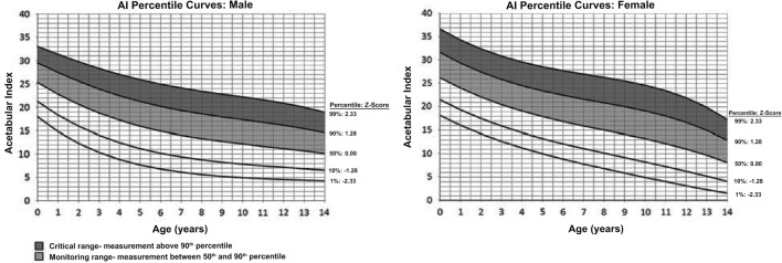
Discussion: Age at the time of initial reduction and the initial severity of DDH have been linked to residual acetabular dysplasia. An anteroposterior pelvic radiograph is the main diagnostic modality, but MRI also provides valuable information, particularly in equivocal cases. The literature supports intervening when significant residual acetabular dysplasia persists at 4-5 years of age, and common surgical indications include acetabular index (AI) > 25°-30°, lateral center-edge angle (LCEA) < 8°-10°, and a broken Shenton's line on radiographs; and a cartilaginous acetabular angle (CAI) > 18°, cartilaginous center-edge angle (CCE) < 13°, and/or the presence of high-signal intensity areas on MRI. Surgical options include redirectional pelvic osteotomies and reshaping acetabuloplasties, which provide comparable radiographic and clinical results.

Conclusion: RAD is common after treatment of DDH and requires regular follow-up for diagnosis and appropriate management to decrease the long-term risk of OA. Long-term outcomes of patients treated with pelvic osteotomies are generally favorable, and the risk of OA can be decreased, although the risk of total hip replacement in the long-term remains.

Keywords: Developmental dysplasia of the hip; Osteoarthritis; Pediatric hip; Pediatrics; Pelvic osteotomy; Residual acetabular dysplasia.


Conflict of interest statement

Conflict of InterestThe authors declare that they have no conflict of interest.

Figures

Fig. 1
Fig. 1
Radiographic (A) and MRI (B) measures of hip dysplasia in a 3-year-old girl with right residual hip dysplasia following a successful initial closed reduction who later underwent an acetabular osteotomy. Coronal proton-density MRI of a different patient illustrating abnormal signal and a break in the fibrocartilaginous acetabular roof (arrow) due to the abnormal joint forces caused by residual hip dysplasia, which is considered a surgical indication by some authors (C). Acetabular index (AI) and cartilaginous acetabular index (CAI) are measured between Hilgenreiner’s line and the tangent to the acetabular sourcil on radiographs or cartilaginous acetabular sourcil on MRI, respectively. Lateral center–edge angle (LCEA) and cartilaginous cente–edge angle (CCE) are measured as the angle between a tangent to the lateral edge of the sourcil and the perpendicular to Hilgenreiner’s line on radiographs and MRI, respectively. Shenton’s line is the imaginary arch connecting the obturator foramen and medial femoral neck
Fig. 2
Fig. 2
Gender-specific percentile curves for acetabular index (AI). Abnormally high AI for age (between 90 and 99th percentiles) is indicated (adapted from Novais, Eduardo N., et al. “Normal percentile reference curves and correlation of acetabular index and acetabular depth ratio in children.” Journal of Pediatric Orthopaedics 38.3 (2018): 163–169, with permission)
Fig. 3
Fig. 3
An algorithm for management of RAD based on the current evidence. DDH: Developmental dysplasia of the hip, RAD: Residual acetabular dysplasia, AP: Anteroposterior, MRI: Magnetic resonance imaging, AI: Acetabular index, LCEA: Lateral center–edge angle, CAI: Cartilaginous acetabular index, CCE: Cartilaginous lateral center–edge angle, HSIA: High-signal intensity, PAO: Periacetabular osteotomy
Fig. 4
Fig. 4
Lateral (A) and medial (B) views of the pelvis illustrating the various osteotomy paths with each technique. (Adapted with permission from Skaggs, David L., and Mininder Kocher. Master Techniques in Orthopaedic Surgery: Pediatrics. Lippincott Williams & Wilkins, 2015. Chapter 16: Dega Acetabuloplasty.)
Fig. 5
Fig. 5
Left hip dislocation is evident on this AP pelvic radiograph of a 7-month-old girl (A), who subsequently underwent a successful closed reduction. Standing AP radiograph at 3 years of age (B) shows residual hip dysplasia, with right AI = 28° (slightly dysplastic) and left AI = 34° (more clearly dysplastic). MRI revealed a right CAI = 10° and CCE = 25° (reassuring) and left CAI = 19° and CCE = 12° (C). A left Dega acetabuloplasty alone (D) was performed following shared decision-making with the family. Radiographs at the age of 6 (e) demonstrating excellent coverage and a class I Severin outcome

References

    1. Wyles CC, Heidenreich MJ, Jeng J, et al. The John Charnley award: redefining the natural history of osteoarthritis in patients with hip dysplasia and impingement. Clinical Orthopaedics and Related Research. 2017;475(2):336–350. - PMC - PubMed
    1. Clohisy JC, Beaulé PE, O'Malley A, et al. Aoa symposium: hip disease in the young adult—current concepts of etiology and surgical treatment. Journal of Bone and Joint Surgery. 2008;90(10):2267–2281. - PubMed
    1. Weinstein SL. Natural history of congenital hip dislocation (cdh) and hip dysplasia. Clinical Orthopaedics and Related Research. 1987;225:62–76. - PubMed
    1. Terjesen T. Residual hip dysplasia as a risk factor for osteoarthritis in 45 years follow-up of late-detected hip dislocation. Journal of Children’s Orthopaedics. 2011;5(6):425–431. - PMC - PubMed
    1. Pun S. Hip dysplasia in the young adult caused by residual childhood and adolescent-onset dysplasia. Current Reviews in Musculoskeletal Medicine. 2016;9(4):427–434. - PMC - PubMed

LinkOut - more resources