. 2021 Dec 28;11(1):e1366.
doi: 10.1002/cti2.1366. eCollection 2022.

Maintained partial protection against Streptococcus pneumoniae despite B-cell depletion in mice vaccinated with a pneumococcal glycoconjugate vaccine

Affiliations

Maintained partial protection against Streptococcus pneumoniae despite B-cell depletion in mice vaccinated with a pneumococcal glycoconjugate vaccine

Giuseppe Ercoli et al. Clin Transl Immunology. .

Abstract

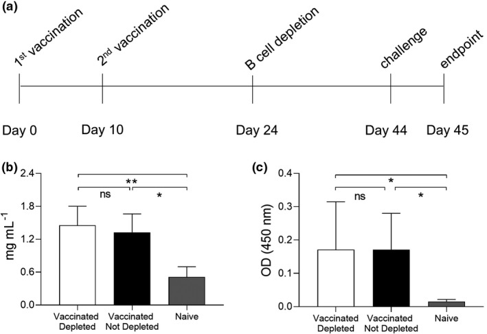
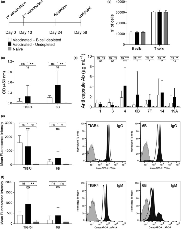
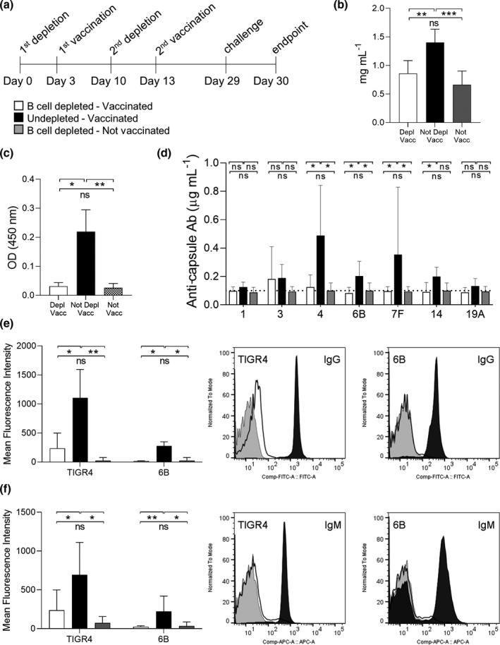
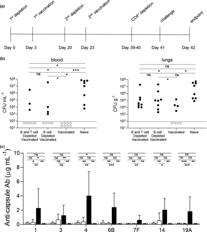
Objectives: Anti-CD20 monoclonal antibody therapy rapidly depletes > 95% of CD20+ B cells from the circulation. B-cell depletion is an effective treatment for autoimmune disease and B-cell malignancies but also increases the risk of respiratory tract infections. This effect on adaptive immunity could be countered by vaccination. We have used mouse models to investigate the effects of B-cell depletion on pneumococcal vaccination, including protection against infection and timing of vaccination in relation to B-cell depletion.

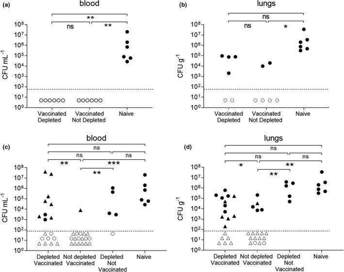
Methods: C57BL/6 female mice were B-cell depleted using anti-CD20 antibody and immunized with two doses of Prevnar-13 vaccine either before or after anti-CD20 treatment. B-cell repertoire and Streptococcus pneumoniae-specific IgG levels were measured using whole-cell ELISA and flow cytometry antibody-binding assay. Protection induced by vaccination was assessed by challenging the mice using a S. pneumoniae pneumonia model.

Results: Antibody responses to S. pneumoniae were largely preserved in mice B-cell depleted after vaccination resulting in full protection against pneumococcal infections. In contrast, mice vaccinated with Prevnar-13 while B cells were depleted (with > 90% reduction in B-cell numbers) had decreased circulating anti-S. pneumoniae IgG and IgM levels (measured using ELISA and flow cytometry antibody binding assays). However, some antibody responses were maintained, and, although vaccine-induced protection against S. pneumoniae infection was impaired, septicaemia was still prevented in 50% of challenged mice.

Conclusions: This study showed that although vaccine efficacy during periods of profound B-cell depletion was impaired some protective efficacy was preserved, suggesting that vaccination remains beneficial.

Keywords: B‐cell depletion; CD20; Streptococcus pneumoniae; immunity; vaccination.


Conflict of interest statement

GW was employed by the company Novartis Institute for BioMedical Research. The remaining authors declare that the research was conducted in the absence of any commercial or financial relationships that could be construed as a potential conflict of interest.

Figures

Figure 1
Figure 1
Vaccination B‐cell depletion. (a) Full schedule of Prevnar‐13 vaccination followed by B‐cell depletion treatment with aCD20 antibody on C57BL/6 mice. B‐cell depletion treatments were administered by intraperitoneal (i.p.) injection of 50 µg of aCD20 antibody, vaccinations by i.p. injection of 20 µL volume of Prevnar‐13 vaccine and challenge by intranasal instillation of 107 pneumococcal colony forming units (CFU). A single mouse experiment was performed with mice divided into three groups depending on the treatment received: vaccinated depleted (n = 6), vaccinated not depleted (n = 6) and naive (n = 6). (b) Total IgG concentrations (mg mL−1) have been measured in mouse sera collected at the endpoint. (c) IgG specific to pneumococcus were also measured for all the mouse sera using a whole‐cell ELISA assay. In b and c , columns represent the average values for each group, error bars indicate Standard Deviations and the Kruskal–Wallis's multiple comparison post hoc was used for statistical analysis (ns, not significant, *P < 0.05 and **P < 0.01). All the sera were tested in triplicate and average values are reported.
Figure 2
Figure 2
Effect of B cell reconstitution on anti‐pneumococcus antibodies. (a) Schedules of the depletion and vaccination treatments extended to allow B cell reconstitution are reported. A single mouse experiment was performed with mice divided into three groups depending on the treatment received: vaccinated depleted (n = 6), vaccinated not depleted (n = 6) and naive (n = 6) (b) Number of B (CD19+) and T cells (CD3+) measured by flow cytometry for 100 000 isolated splenocytes are shown. In all histograms, columns represent average values and error bars standard deviations, depleted‐vaccinated mice are shown in white, not depleted‐vaccinated in black and naïve in grey. (c) Mouse sera collected at the endpoint were used to measure IgG specific to TIGR4 and 6B pneumococcal strains by whole‐cell ELISA assay. (d) Meso Scale Discovery (MSD) assay was used to measure the specific IgG response to seven pneumococcal capsule serotypes by quantifying their concentration (µg mL−1). The dotted line indicates the limit of detection. (e, f) Serum deposition assay was used to measure the amount of surface‐exposed pneumococcal‐specific IgG (e) and IgM (f). The average mean fluorescence intensity (MFI) for each group is reported in the left panels. Representative histograms showing a shift in populations with positive antibody binding against TIGR4 and 6B pneumococcal strains are shown in the right panels. Depleted (white), not depleted (black) and not vaccinated (grey) representative samples are shown. For all experiments, the sera were tested in triplicate and average values are reported; the Kruskal–Wallis test followed by Dunn's multiple comparison post hoc was used for statistical analysis (ns, not significant, *P < 0.05; ** and P < 0.01). Representative histograms showing a shift in populations positive for IgM against TIGR4 and 6B pneumococcal strains are shown in the right panels.
Figure 3
Figure 3
Effects of B‐cell depletion before Prevnar‐13 vaccination on serological responses. (a) Full schedule of B‐cell depletion treatment with aCD20 antibody followed by Prevnar‐13 vaccination on C57BL/6 mice. A single mouse experiment was performed with mice divided into three groups depending on the treatment received: depleted vaccinated (n = 6), not depleted vaccinated (n = 6) and not vaccinated (n = 6). (b) Total IgG concentrations (mg mL−1) in sera collected at the endpoint have been measured for each group. In all histograms, columns represent average values and error bars standard deviations, depleted‐vaccinated mice are shown in white, not depleted‐vaccinated in black and not vaccinated in grey. (c) IgG specific to pneumococcus have been measured for all the mouse sera using a whole‐cell ELISA assay against Streptococcus pneumoniae strain 6B. (d) The Meso Scale Discovery (MSD) assay was used to measure the specific IgG response to seven pneumococcal capsule serotypes by quantifying their concentration (µg mL−1). The dotted line indicates the limit of detection. (e, f) Serum deposition assay was used to measure the amount of surface‐exposed pneumococcal‐specific IgG (e) and IgM (f). The average mean fluorescence intensity for each group is reported in the left panels. Representative histograms showing a shift in populations with positive antibody binding against TIGR4 and 6B pneumococcal strains are shown in the right panels. Depleted (white), not depleted (black) and not vaccinated (grey) representative samples are shown. For all experiments, the sera were tested in triplicate and average values are reported, the Kruskal–Wallis test followed by Dunn's multiple comparison post hoc was used for statistical analysis (ns, not significant, *P < 0.05; **P < 0.01 and ***P < 0.001). Representative histograms showing a shift in populations positive for IgM against TIGR4 and 6B pneumococcal strains are shown in the right panels.
Figure 4
Figure 4
Effects of B‐cell depletion on IP Prevnar‐13 vaccination–induced protection determined after pneumonia challenge with strain Streptococcus pneumoniae 6B strain BHN418. In the vaccination‐depletion study pneumococcal pneumonia was induced by infecting the mice intranasally with the 6B strain at day 44. (a, b) A single mouse experiment was performed with mice divided into three groups depending on the treatment received: vaccinated depleted (n = 6), vaccinated not depleted (n = 6) and naive (n = 6). Twenty‐four hours after the infection, colony forming units (CFU) were measured from blood (a) and lungs (b). In the depletion‐vaccination study pneumococcal pneumonia was induced by infecting the mice intranasally with 6B strain at day 29. (c, d) Mice were divided in 4 groups depending on the treatment received: B‐cell–depleted vaccinated (n = 18), not depleted vaccinated (n = 18) depleted not vaccinated naïve (n = 5) and naïve (n = 6). Data from two independent experiments were pooled, with experiment 1 represented by circular symbols and experiment 2 (with no control groups) triangular symbols. Twenty‐four hours after the infection, CFU were measured from blood (c) and lungs (d). Each symbol represents the CFU value from a single mouse; mice were considered infected (black dots) when their CFU number was higher than the limit of detection (dotted line). Empty dots represent uninfected mice with 0 recovered CFU. Their position on the Y axis is varied to improve visual presentation and does not represent any differences in CFU. Statistical differences between groups are indicated by thin black lines (ns, not significant, *P < 0.05 and **P < 0.01 and ***P < 0.001. Fisher’s exact test, two‐tailed).
Figure 5
Figure 5
Effects of IM vaccination on protection against Streptococcus pneumoniae 6B strain BHN418 pneumonia challenge. (a) Pneumococcal pneumonia was induced infecting the mice intranasally with the 6B strain at day 41 after two rounds of intramuscular Prevnar‐13 vaccination. A single mouse experiment was performed with mice divided into four groups depending on the treatment received: B‐cell depleted then vaccinated and given additional T‐cell depletion pre‐challenge (n = 8), B‐cell depleted then vaccinated (n = 8), vaccinated (n = 8) and naïve unvaccinated (n = 8). Twenty‐four hours after the infection, colony forming units (CFU) were measured from blood and lungs (b). Each symbol represents the CFU value from a single mouse; mice were considered infected (black dots) when their CFU number was higher than the limit of detection (dotted line). Empty dots represent uninfected mice with 0 recovered CFU. Statistical differences between groups are indicated by thin black lines (ns, not significant, *P < 0.05 and ***P < 0.001. Fisher's exact test, two‐tailed). (c) The Meso Scale Discovery (MSD) assay was used to measure the specific IgG response to seven pneumococcal capsule serotypes by quantifying their concentration (µg mL−1). The sera were tested in triplicate, and average values are reported. The dotted line indicates the limit of detection. The Kruskal–Wallis test followed by Dunn's multiple comparison post hoc was used for statistical analysis (ns, not significant, *P < 0.05; **P < 0.01 and ***P < 0.001).

Similar articles

Cited by

References

    1. Kurosaki T, Kometani K, Ise W. Memory B cells. Nat Rev Immunol 2015; 15: 149–159. - PubMed
    1. Rodriguez‐Pinto D. B cells as antigen presenting cells. Cell Immunol 2005; 238: 67–75. - PubMed
    1. Mauri C, Menon M. Human regulatory B cells in health and disease: therapeutic potential. J Clin Invest 2017; 127: 772–779. - PMC - PubMed
    1. Jonas C. Rituxan: the new kid on the block. Oncol Nurs Forum 1998; 25: 669. - PubMed
    1. Rubbert‐Roth A, Tak PP, Zerbini C et al. Efficacy and safety of various repeat treatment dosing regimens of rituximab in patients with active rheumatoid arthritis: results of a Phase III randomized study (MIRROR). Rheumatology (Oxford) 2010; 49: 1683–1693. - PMC - PubMed