

Expression of Otx Genes in Müller Cells Using an In Vitro Experimental Model of Retinal Hypoxia
- PMID: 35003791
- PMCID: PMC8741358
- DOI: 10.1155/2021/6265553
Expression of Otx Genes in Müller Cells Using an In Vitro Experimental Model of Retinal Hypoxia
Abstract
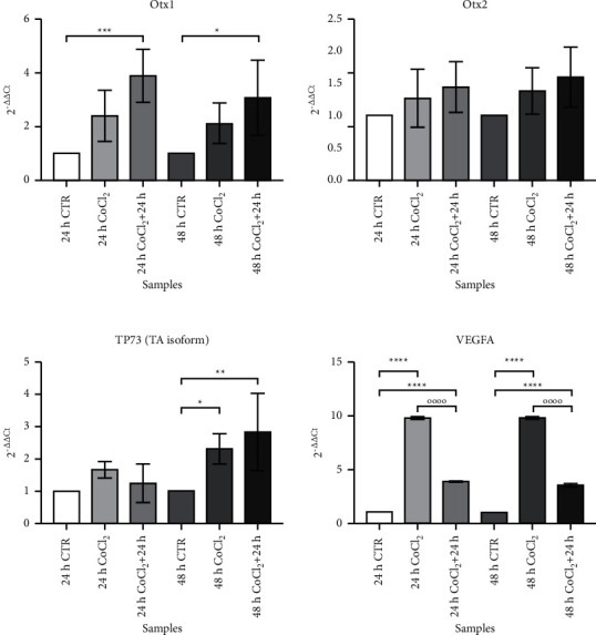
Introduction: Müller glial cells typically activate to react to hypoxic tissue damage in several retinal diseases. We evaluated the in vitro response to a hypoxia-mimicking stimulus on the expression of a set of genes, known to contribute to eye morphogenesis and cell differentiation.
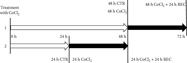
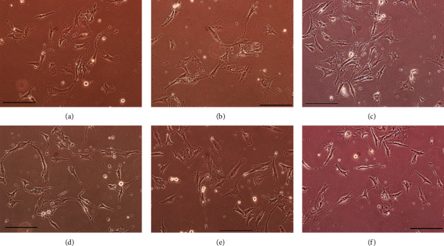
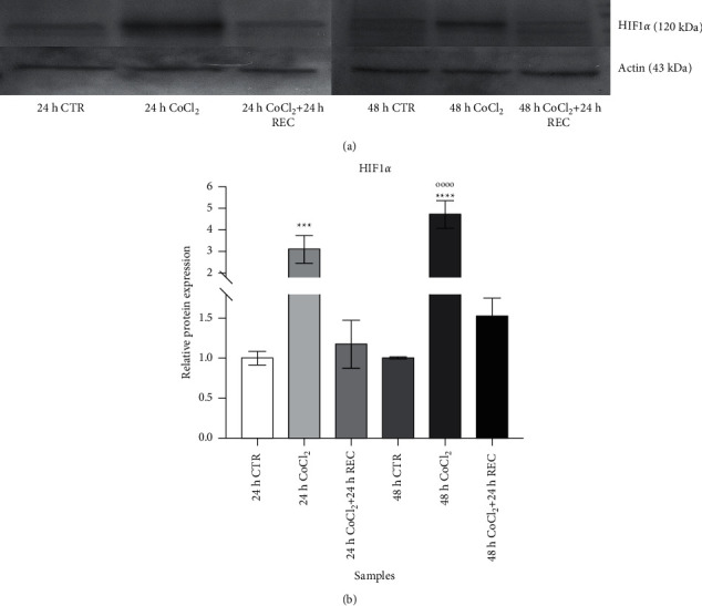
Materials and methods: A MIO-M1 Müller cell line was cultured in a hypoxia-mimicking environment by the addition of cobalt chloride to the culture medium, followed by a recovery time in which we mimic restoration from the hypoxic insult. The HIF-1α protein and VEGF-A gene expression were quantified to verify the induction of a hypoxia-like state.
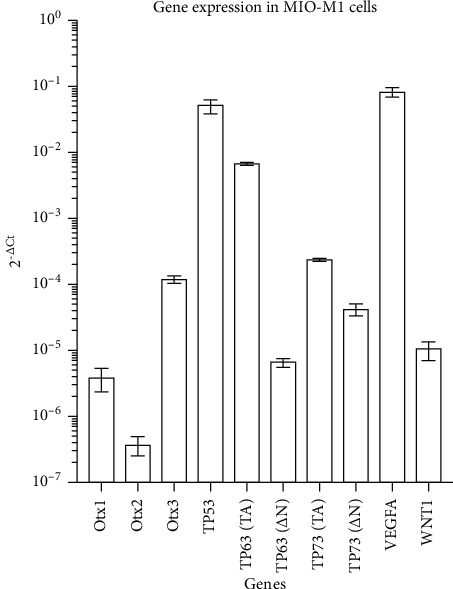
Results: Among the genes under study, we did not observe any difference in the expression levels of Otx1 and Otx2 during treatment; conversely, Otx1 was overexpressed during recovery steps. The VEGF-A gene was strongly upregulated at both the CoCl2 and recovery time points. The transactivated isoform (TA) of the TP73 gene showed an overexpression in long-term exposure to the hypoxic stimulus with a further increase after recovery. Discussion. Our molecular analysis is able to describe the activation of a set of genes, never before described, that can drive the response to a hypoxia-like status. The improved comprehension of these cellular events will be useful for designing new therapeutical approaches for retinal pathologies.
Copyright © 2021 Claudio Azzolini et al.
Conflict of interest statement
The authors declare no conflicts of interest.
Figures
References
-
- Willbold E., Layer P. G. Müller glia cells and their possible roles during retina differentiation in vivo and in vitro. Histology & Histopathology . 1998;13(2):531–552. - PubMed
LinkOut - more resources
Full Text Sources
