

Cobb's Tufts: A Systematic Review
- PMID: 35003982
- PMCID: PMC8723767
- DOI: 10.7759/cureus.20151
Cobb's Tufts: A Systematic Review
Abstract
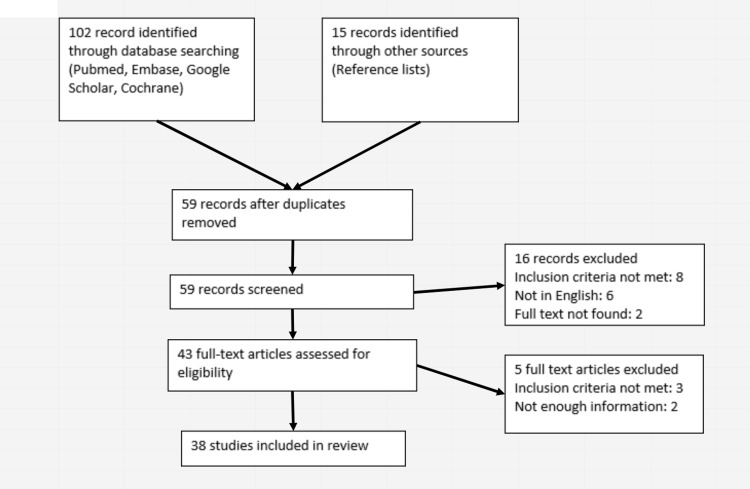
Cobb's tufts, also known as iris vascular tufts (IVT) and iris microhemangiomas (IMH), are coils of tightly clustered, minute blood vessels at the iris pupillary border. This study aimed to analyze previous literature and provide an update on Cobb's tufts. A systematic literature review was carried out by interrogating PubMed, Google Scholar, Cochrane, and Embase databases. Full-text English language articles of any year were included in this study. A total of 38 articles fulfilled our inclusion criteria. A total of 115 reported cases of Cobb's tufts were incorporated into our review. The age of the patients ranged between 36 and 86 years. No sex or racial predisposition was noted. Most patients had no history of trauma, surgery, or blood dyscrasia. The majority of cases are asymptomatic and bilateral unless a spontaneous hyphema occurs, which most commonly presents as blurred vision. The etiology of this condition remains uncertain; however, a higher incidence has been shown in systemic conditions such as myotonic dystrophy and diabetes. Fluorescein angiography can be utilized to investigate tufts. Management includes treatment of raised intraocular pressure, observation for single bleeds, laser therapy for recurrent hyphemas, and lastly, iridectomy, which is considered in cases of recurrence following laser treatment.
Keywords: cobb's tufts; hyphema; iris microhaemangioma; iris vascular tufts; spontaneous hyphema.
Copyright © 2021, Almafreji et al.
Conflict of interest statement
The authors have declared that no competing interests exist.
Figures
Similar articles
-
Iris microhaemangioma: a management strategy.Int J Ophthalmol. 2013 Apr 18;6(2):246-50. doi: 10.3980/j.issn.2222-3959.2013.02.26. Print 2013. Int J Ophthalmol. 2013. PMID: 23638431 Free PMC article.
-
Iris vascular tuft causing recurrent hyphema and raised IOP: a new indication for laser photocoagulation, angiographic follow-up, and review of laser outcomes.J Glaucoma. 2010 Jun-Jul;19(5):336-8. doi: 10.1097/IJG.0b013e3181bd899b. J Glaucoma. 2010. PMID: 19855294
-
Cobb's tufts: a rare cause of spontaneous hyphaema.Int Ophthalmol. 2001;24(6):299-300. doi: 10.1023/b:inte.0000006761.23314.22. Int Ophthalmol. 2001. PMID: 14750565
-
Medical interventions for traumatic hyphema.Cochrane Database Syst Rev. 2019 Jan 14;1(1):CD005431. doi: 10.1002/14651858.CD005431.pub4. Cochrane Database Syst Rev. 2019. Update in: Cochrane Database Syst Rev. 2023 Mar 13;3:CD005431. doi: 10.1002/14651858.CD005431.pub5. PMID: 30640411 Free PMC article. Updated.
-
Medical interventions for traumatic hyphema.Cochrane Database Syst Rev. 2023 Mar 13;3(3):CD005431. doi: 10.1002/14651858.CD005431.pub5. Cochrane Database Syst Rev. 2023. PMID: 36912744 Free PMC article. Review.
Cited by
-
Oral tranexamic acid for acute management of active bleeding from iris microhemangiomatosis: A case report.Am J Ophthalmol Case Rep. 2024 Jan 19;33:102000. doi: 10.1016/j.ajoc.2024.102000. eCollection 2024 Mar. Am J Ophthalmol Case Rep. 2024. PMID: 38318444 Free PMC article.
-
Spontaneous hyphema from iris microhemangioma in Eisenmenger syndrome.Am J Ophthalmol Case Rep. 2022 Apr 12;26:101536. doi: 10.1016/j.ajoc.2022.101536. eCollection 2022 Jun. Am J Ophthalmol Case Rep. 2022. PMID: 35496761 Free PMC article.
References
-
- Vascular tufts at the pupillary margin: a preliminary report on 44 patients. Cobb B. https://pubmed.ncbi.nlm.nih.gov/5272254/ Trans Ophthalmol Soc U K. 1969;88:211–221. - PubMed
-
- Vascular tufts at the pupillary margin in myotonic dystrophy. Cobb B, Shilling JS, Chisholm IH. Am J Ophthalmol. 1970;69:573–582. - PubMed
Publication types
LinkOut - more resources
Full Text Sources