Post-chemotherapy Changes in Bone Marrow in Acute Leukemia With Emphasis on Detection of Residual Disease by Immunohistochemistry
- PMID: 35004001
- PMCID: PMC8725173
- DOI: 10.7759/cureus.20175
Post-chemotherapy Changes in Bone Marrow in Acute Leukemia With Emphasis on Detection of Residual Disease by Immunohistochemistry
Abstract
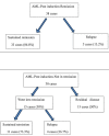
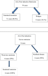
Introduction In acute leukemia, the leading cause of treatment failure is disease relapse leading to a low level of complete remission and short overall survival. Post-chemotherapy marrow examination gives vital clues regarding treatment response and marrow regeneration. Aim We aimed to study the histomorphological changes in post-chemotherapy bone marrow in acute leukemias, monitor residual disease by immunohistochemistry (IHC) on trephine biopsy, and correlate survival status. Method This study was a prospective clinical study. A total of 155 post-induction cases (acute myeloid leukemia [AML] - 68 and acute lymphoblastic leukemia [ALL] - 87), from January 2014 to December 2015, were included with a follow-up of 4-28 months. A detailed histomorphology was studied in all cases. IHC was applied in 88 cases of post-induction marrow, which showed morphologic suspicion of an increase in blasts. Observations Post-induction marrow was hypercellular in 55.9% of AML and normocellular in 56.3% of ALL. Regenerative hematopoiesis was noted in 37.4% of AML and 88.5% of cases of ALL. Marrow serous atrophy and stromal edema were associated with delayed recovery of counts and their recovery duration ranged from one to five months. Twenty-seven bone marrow aspirates were unsatisfactory, and their trephine biopsies were showed remission in 20 cases and stromal changes in nine cases. In addition, trephine biopsy picked up residual leukemic blasts in four cases in which aspirate showed remission status. Post-induction marrow IHC with scattered positivity for blasts showed sustained remission in 96% cases, and in those with clustered positivity, 28.6% showed residual disease, and 7.2% showed relapse at the end of the study period. The median survival duration was 13, 3, and 12 months for cases with sustained remission, residual disease, and relapse, respectively. There was a statistically significant difference in median survival of patients in the three groups (sustained remission, residual disease, and relapse) (p=0.000). Conclusion We conclude that histomorphology augmented by IHC on trephine biopsy gives valuable information regarding post-chemotherapy changes and residual disease status. Bone marrow trephine biopsy is an important tool to assess the remission status of patients with acute leukemia.
Keywords: acute leukemia; bone marrow regeneration; dyspoiesis; histomorphology of bone marrow; post-induction chemotherapy; remission; residual disease; serous atrophy; stromal changes; trephine biopsy.
Copyright © 2021, Ayyanar et al.
Conflict of interest statement
The authors have declared that no competing interests exist.
Figures


References
-
- Clustered immature myeloid precursors in intertrabecular region during remission evolve from leukemia stem cell near endosteum and contribute to disease relapse in acute myeloid leukemia. Wu Z, Yu Y, Zhang J, Zhai Y, Tao Y, Shi J. Med Hypotheses. 2013;80:624–628. - PubMed
-
- Evaluation of post chemotherapy bone marrow changes in acute Leukaemia. Belurkar S, Nepali PB, Manandhar B, et al. http://www.ijsrp.org/research-paper-0215/ijsrp-p3813 Int J Sci Res Publ. 2015;5:2250–3153.
-
- Immunophenotypic analysis of hematogones (B-lymphocyte precursors) in 662 consecutive bone marrow specimens by 4-color flow cytometry. McKenna RW, Washington LT, Aquino DB, Picker LJ, Kroft SH. Blood. 2001;98:2498–2507. - PubMed
LinkOut - more resources
Full Text Sources
