

Response of Pediatric Choroidal Neovascularization to Anti-Vascular Endothelial Growth Factor
- PMID: 35004017
- PMCID: PMC8727322
- DOI: 10.7759/cureus.20195
Response of Pediatric Choroidal Neovascularization to Anti-Vascular Endothelial Growth Factor
Abstract
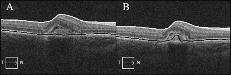
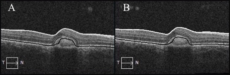
Choroidal neovascularization (CNV) is a rare condition in children but poses a substantial threat to vision. Anti-vascular endothelial growth factor (anti-VEGF) therapy is commonly used in the pediatric population to treat retinopathy of prematurity. However, the use of anti-VEGF is less common for childhood CNV due to the rarity of CNV in the pediatric population. We report the case of a 10-year-old male presenting with an idiopathic choroidal neovascular membrane. Following a relapse of subretinal fluid after photodynamic therapy, anti-VEGF (bevacizumab) was injected and resulted in remission of the neovascular membrane and improved visual outcome. Further studies are required to elucidate the long-term outcomes associated with the use of anti-VEGF in pediatric patients.
Keywords: anti-vegf; choroidal neovascular membrane; ophthalmology; pediatric ophthalmology; retina.
Copyright © 2021, Ruparelia et al.
Conflict of interest statement
The authors have declared that no competing interests exist.
Figures


References
-
- Anti-vascular endothelial growth factor indications in ocular disease. Cornel S, Adriana ID, Mihaela TC, Speranta S, Algerino S, Mehdi B, Jalaladin HR. https://pubmed.ncbi.nlm.nih.gov/29450313/ Rom J Ophthalmol. 2015;59:235–242. - PMC - PubMed
-
- Management of pediatric choroidal neovascular membranes with intravitreal anti-VEGF agents: a retrospective consecutive case series. Kohly RP, Muni RH, Kertes PJ, Lam WC. Can J Ophthalmol. 2011;46:46–50. - PubMed
Publication types
LinkOut - more resources
Full Text Sources