Review
. 2021 Dec 22:9:756930.
doi: 10.3389/fbioe.2021.756930. eCollection 2021.

Tackling the Challenges of Graft Healing After Anterior Cruciate Ligament Reconstruction-Thinking From the Endpoint

Affiliations
Review

Tackling the Challenges of Graft Healing After Anterior Cruciate Ligament Reconstruction-Thinking From the Endpoint

Shiyi Yao et al. Front Bioeng Biotechnol. .

Abstract

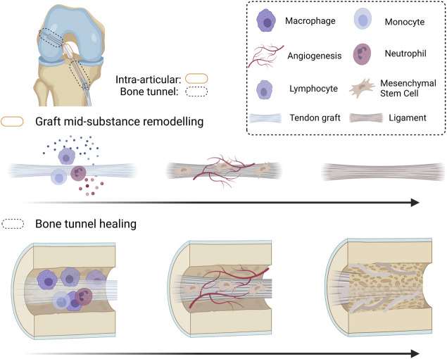
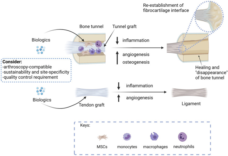
Anterior cruciate ligament (ACL) tear is common in sports and accidents, and accounts for over 50% of all knee injuries. ACL reconstruction (ACLR) is commonly indicated to restore the knee stability, prevent anterior-posterior translation, and reduce the risk of developing post-traumatic osteoarthritis. However, the outcome of biological graft healing is not satisfactory with graft failure after ACLR. Tendon graft-to-bone tunnel healing and graft mid-substance remodeling are two key challenges of biological graft healing after ACLR. Mounting evidence supports excessive inflammation due to ACL injury and ACLR, and tendon graft-to-bone tunnel motion negatively influences these two key processes. To tackle the problem of biological graft healing, we believe that an inductive approach should be adopted, starting from the endpoint that we expected after ACLR, even though the results may not be achievable at present, followed by developing clinically practical strategies to achieve this ultimate goal. We believe that mineralization of tunnel graft and ligamentization of graft mid-substance to restore the ultrastructure and anatomy of the original ACL are the ultimate targets of ACLR. Hence, strategies that are osteoinductive, angiogenic, or anti-inflammatory should drive graft healing toward the targets. This paper reviews pre-clinical and clinical literature supporting this claim and the role of inflammation in negatively influencing graft healing. The practical considerations when developing a biological therapy to promote ACLR for future clinical translation are also discussed.

Keywords: ACL; ACL reconstruction; angiogenesis; anterior cruciate ligament; biological therapy; graft healing; inflammation; osteogenesis.


Conflict of interest statement

The authors declare that the research was conducted in the absence of any commercial or financial relationships that could be construed as a potential conflict of interest.

Figures

FIGURE 1
FIGURE 1
Schematic diagram summarizing the graft healing process after anterior cruciate ligament (ACL) reconstruction. After ACL reconstruction (ACLR), inflammatory occurs, attracting immune cells and mesenchymal stromal cells (MSCs) to the injured site. The original cells in the tendon graft undergo necrosis and are replaced by MSCs infiltrating into the graft. Both MSCs and the inflammatory cells produce angiogenic factors and the MSCs proliferate and differentiate. The differentiated MSCs produce extracellular matrix and remodeling enzymes to incorporate the tendon graft-to-bone tunnel by Sharpey’s fibers and is associated with improved biomechanical properties of the healing complex. However, there is regional variation in healing along the bone tunnel. The original ACL insertion site is not re-established. The tendon graft mid-substance theoretically should remodel to a ligament. However, it degenerates due to excessive inflammation and poor angiogenesis after ACLR. Created with BioRender.com.
FIGURE 2
FIGURE 2
A summary of the treatment approaches and considerations for developing a clinically viable biological therapy for the promotion of graft healing after ACLR. The ultimate goal of ACLR is to replace the tunnel graft by bone with re-establishment of direct insertion at the original ACL footprint and remodel the tendon graft mid-substances to a ligament to meet the functions of an ACL. Biologics that promote bone healing, therefore, should promote tunnel healing, while biologics that enhance angiogenesis are expected to accelerate both tunnel healing and graft remodeling. As inflammation hampers graft healing, biologics that suppress inflammation are anticipated to promote both tunnel healing and graft remodeling. Researches developing osteogenic, angiogenic, or anti-inflammatory biologicals should consider if the proposed intervention is arthroscopy compatible, supports sustainable and site-specific application, and meets quality control requirements for successful future clinical translation. Created with BioRender.com.

Similar articles

Cited by

References

    1. Amiel D., Frank C., Harwood F., Fronek J., Akeson W. (1983). Tendons and Ligaments: a Morphological and Biochemical Comparison. J. Orthop. Res. 1 (3), 257–265. 10.1002/jor.1100010305 - DOI - PubMed
    1. Anderson K., Seneviratne A. M., Izawa K., Atkinson B. L., Potter H. G., Rodeo S. A. (2001). Augmentation of Tendon Healing in an Intraarticular Bone Tunnel with Use of a Bone Growth Factor. Am. J. Sports Med. 29 (6), 689–698. 10.1177/03635465010290060301 - DOI - PubMed
    1. Berdis A. S., Veale K., Fleissner P. R., Jr. (2019). Outcomes of Anterior Cruciate Ligament Reconstruction Using Biologic Augmentation in Patients 21 Years of Age and Younger. Arthrosc. J. Arthroscopic Relat. Surg. 35 (11), 3107–3113. 10.1016/j.arthro.2019.05.047 - DOI - PubMed
    1. Berg E. E., Pollard M. E., Kang Q. (2001). Interarticular Bone Tunnel Healing. Arthrosc. J. Arthroscopic Relat. Surg. 17 (2), 189–195. 10.1053/jars.2001.20958 - DOI - PubMed
    1. Brophy R. H., Kovacevic D., Imhauser C. W., Stasiak M., Bedi A., Fox A. J., et al. (2011). Effect of Short-Duration Low-Magnitude Cyclic Loading versus Immobilization on Tendon-Bone Healing after ACL Reconstruction in a Rat Model. The J. Bone Jt. Surgery-American Volume 93 (4), 381–393. 10.2106/jbjs.I.00933 - DOI - PMC - PubMed

LinkOut - more resources