Review
. 2021 Dec 23:9:787551.
doi: 10.3389/fbioe.2021.787551. eCollection 2021.

Technological Approaches in the Analysis of Extracellular Vesicle Nucleotide Sequences

Affiliations
Review

Technological Approaches in the Analysis of Extracellular Vesicle Nucleotide Sequences

Tine Tesovnik et al. Front Bioeng Biotechnol. .

Abstract

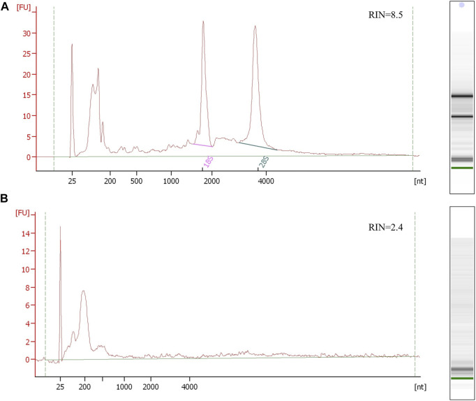
Together with metabolites, proteins, and lipid components, the EV cargo consists of DNA and RNA nucleotide sequence species, which are part of the intracellular communication network regulating specific cellular processes and provoking distinct target cell responses. The extracellular vesicle (EV) nucleotide sequence cargo molecules are often investigated in association with a particular pathology and may provide an insight into the physiological and pathological processes in hard-to-access organs and tissues. The diversity and biological function of EV nucleotide sequences are distinct regarding EV subgroups and differ in tissue- and cell-released EVs. EV DNA is present mainly in apoptotic bodies, while there are different species of EV RNAs in all subgroups of EVs. A limited sample volume of unique human liquid biopsy provides a small amount of EVs with limited isolated DNA and RNA, which can be a challenging factor for EV nucleotide sequence analysis, while the additional difficulty is technical variability of molecular nucleotide detection. Every EV study is challenged with its first step of the EV isolation procedure, which determines the EV's purity, yield, and diameter range and has an impact on the EV's downstream analysis with a significant impact on the final result. The gold standard EV isolation procedure with ultracentrifugation provides a low output and not highly pure isolated EVs, while modern techniques increase EV's yield and purity. Different EV DNA and RNA detection techniques include the PCR procedure for nucleotide sequence replication of the molecules of interest, which can undergo a small-input EV DNA or RNA material. The nucleotide sequence detection approaches with their advantages and disadvantages should be considered to appropriately address the study problem and to extract specific EV nucleotide sequence information with the detection using qPCR or next-generation sequencing. Advanced next-generation sequencing techniques allow the detection of total EV genomic or transcriptomic data even at the single-molecule resolution and thus, offering a sensitive and accurate EV DNA or RNA biomarker detection. Additionally, with the processes where the EV genomic or transcriptomic data profiles are compared to identify characteristic EV differences in specific conditions, novel biomarkers could be discovered. Therefore, a suitable differential expression analysis is crucial to define the EV DNA or RNA differences between conditions under investigation. Further bioinformatics analysis can predict molecular cell targets and identify targeted and affected cellular pathways. The prediction target tools with functional studies are essential to help specify the role of the investigated EV-targeted nucleotide sequences in health and disease and support further development of EV-related therapeutics. This review will discuss the biological diversity of human liquid biopsy-obtained EV nucleotide sequences DNA and RNA species reported as potential biomarkers in health and disease and methodological principles of their detection, from human liquid biopsy EV isolation, EV nucleotide sequence extraction, techniques for their detection, and their cell target prediction.

Keywords: DNA; RNA; biomarkers; extracellular vesicles (EVs); nucleotide sequences detection; therapeutics.


Conflict of interest statement

The authors declare that the research was conducted in the absence of any commercial or financial relationships that could be construed as a potential conflict of interest.

Figures

FIGURE 1
FIGURE 1
EV isolation procedure. The scheme shows the procedure for blood plasma EV isolation from the first steps after the blood collection. The samples undergo centrifugation steps to separate plasma and blood cells, while the next centrifugation step is used to remove cell debris and large protein complexes. The samples can be stored with deep freezing or further processed with methods for EV isolation. EVs can be isolated by (A) ultracentrifugation at high speeds, using (B) precipitation reagent and separated by low-speed centrifugation, (C) immunoprecipitation with antibodies on magnetic beads in the magnetic field, or (D) size exclusion chromatography based on the particle size separation on the column.
FIGURE 2
FIGURE 2
Electropherogram profiles of kidney and urine PEG-isolated EV RNA samples using the Bioanalyzer Pico 6,000 kit. (A) The upper electropherogram RNA profile of the kidney tissue shows the preserved RNA sample with detectable 16S and 28S rRNA and RNA integrity number (RIN) representing non-degraded RNA. (B) The bottom electropherogram shows urine EV sample RNA, where EVs were isolated by PEG. The RNA profile presents typical EV short RNA lengths and undetectable rRNA peaks, with unreliable sample integrity RIN due to undetectable rRNA. Both RNA samples were isolated using trizol/chloroform extraction (Qiazol, Qiagen) and commercial RNA isolation and purification columns (Qiagen RNeasy kit, Qiagen).
FIGURE 3
FIGURE 3
Two principles of commonly used miRNA qPCR detection. Stem-loop miRNA qPCR uses stem-loop oligos, which align on the miRNA and form the cDNA product in the step of reverse transcription. The miRNA transcripts are then amplified and detected by measuring fluorescence after the reporting probe degradation in the qPCR reaction. The stem-loop miRNA detection has a limited ability of multiplexing, requires multiple transcription reactions in which more miRNA species are detected. Universal miRNA qPCR preparation procedure transcribe all miRNA into cDNA after poly-adenylation and universal oligo ligation. The selected miRNAs are detected with specific probes capable of detecting different miRNA in multiplex reactions from the same transcription reaction.
FIGURE 4
FIGURE 4
EV miRNA NGS library quality control and sequencing report. (A) Electropherogram of the plasma EV small RNA NGS library, with peaks of adapter dimers (120 nt), incorporated miRNA fragments (143 nt), and larger RNA inserts (194 nt). The NGS library was prepared using the NEBNext small RNA library kit and after 50cycle single-end Illumina sequencing, fastq files were generated. Sequencing data were analyzed using the sRNAtoolbox—a collection of small RNA analysis tools, where sequencing data were trimmed for NGS adapters, aligned on the human regerence genome and annotated with miRBase miRNA reference sequence data. Graphical presentation of the aligned reads is shown on graph with (B) read length distribution and (C) RNA length distribution by non-coding RNA species.
FIGURE 5
FIGURE 5
Scheme of the miRNA NGS data analysis from data generation to the final result. The Illumina sequencing platform generates bcl files, which are translated in the process of base-calling and demultiplexing by the sample index to fastq files for each studied sample. The fastq file includes sequencing run information and nucleotide sequence data with read quality data per nucleotide. In the first step of the data analysis, fastq files of the studied samples undergo the adapter and low quality sequence removal and sequence alignment on a human genome or sequences in databases follows. After alignment, annotation and quantification form a sample expression miRNA profile with the sequencing and read quality report of the studied samples. The differences in studied conditions can be identified with differential expression analysis where samples of studied conditions are compared after the expression normalization, comparison, and statistical evaluation. Final analysis results are represented in tables of normalized identified sequences, tables of differential expression with the statistical comparison, and graphical representation of read distribution and differential expression analysis results.
FIGURE 6
FIGURE 6
Differentially expressed EV miRNA functional study. Differentially expressed miRNAs are exposed by means of synthetic vesicles (DOTAP) to the whole blood cells of the immune system where vesicles with miRNAs are accumulated in the endolysosomal pathway of phagocytes. Vesicle-delivered miRNA overnight incubation can result in an increased expression of activation markers on effector T-cells detected using flow cytometry. The scheme is summarized work of Tesovnik et al. (2020).
FIGURE 7
FIGURE 7
Graphical presentation of basic steps and possible variability factors in the EV processing and EV nucleotide sequence analysis. The human EV design includes the studied group participants’ selection, where biological variability factors play the main including/excluding factors for homogeneous group selection to study specific human physiological conditions. After studying the individuals’ selection, liquid biopsy samples are collected in sterile tubes or containers and processed to a cell-free liquid biopsy medium. Cells and cell debris with larger protein complexes are removed using centrifugation or filtration steps and processed samples are deep-freeze stored before further processing. EVs can be isolated using different isolation techniques and procedures, which can include protein and non-EV nucleotide sequence degradation and yield with different amounts of isolated EVs. Next, EV DNA or RNA are isolated where the nucleotide sequence profile results in a low amount of total EV DNA or RNA with length below 200 nucleotides. Isolated EV nucleotide sequences can be analyzed by qPCR, microarray, or NGS. All of the nucleotide sequence detection techniques have their advantages and disadvantages, while the limitations of all of the methods are the consequence of sequence variability and technical variation in ligation, amplification, and nucleotide pairing with complementary sequences. After the nucleotide sequence detection, the data are analyzed using bioinformatics pipelines and bioinformatics tools. A slightly more demanding analysis is required for NGS data analysis, where bioinformatics analysis parameters with the adapter trimming and sequence annotation step can have a tremendous effect on final differential expression results where data are normalized and differentially expressed EV nucleotide sequences in compared conditions are detected based on the limit parameters. Further bioinformatics data prediction analysis with databases’ information and appropriate variable thresholds can reveal the affected cellular targets, cellular pathways, and EV nucleotide sequences as therapeutic targets, which can be further characterized and confirmed within in vitro or/and in vivo studies. Differentially expressed EVs are also potential biomarkers, while their diagnostic potential must be confirmed with additional experiments on larger cohorts and validation steps.

References

    1. Agarwal V., Bell G. W., Nam J.-W., Bartel D. P. (2015). Predicting Effective microRNA Target Sites in Mammalian mRNAs. Elife 4, e05005. 10.7554/eLife.05005 - DOI - PMC - PubMed
    1. Ahmadi Badi S., Moshiri A., Fateh A., Rahimi Jamnani F., Sarshar M., Vaziri F., et al. (2017). Microbiota-Derived Extracellular Vesicles as New Systemic Regulators. Front. Microbiol. 8, 1610. 10.3389/fmicb.2017.01610 - DOI - PMC - PubMed
    1. Albanese M., Chen Y.-F. A., Hüls C., Gärtner K., Tagawa T., Mejias-Perez E., et al. (2020). Micro RNAs Are Minor Constituents of Extracellular Vesicles and Are Hardly Delivered to Target Cells. bioRxiv [Epub ahead f print]. 10.1101/2020.05.20.106393 - DOI - PMC - PubMed
    1. Alexiou A., Zisis D., Kavakiotis I., Miliotis M., Koussounadis A., Karagkouni D., et al. (2020). DIANA-mAP: Analyzing miRNA from Raw NGS Data to Quantification. Genes 12, 46. 10.3390/genes12010046 - DOI - PMC - PubMed
    1. Ali N., Rampazzo R. d. C. P., Costa A. D. T., Krieger M. A. (2017). Current Nucleic Acid Extraction Methods and Their Implications to Point-of-Care Diagnostics. Biomed. Res. Int. 2017, 1–13. 10.1155/2017/9306564 - DOI - PMC - PubMed

LinkOut - more resources