

Myeloid-Derived Suppressor Cells: A Multifaceted Accomplice in Tumor Progression
- PMID: 35004667
- PMCID: PMC8733653
- DOI: 10.3389/fcell.2021.740827
Myeloid-Derived Suppressor Cells: A Multifaceted Accomplice in Tumor Progression
Abstract
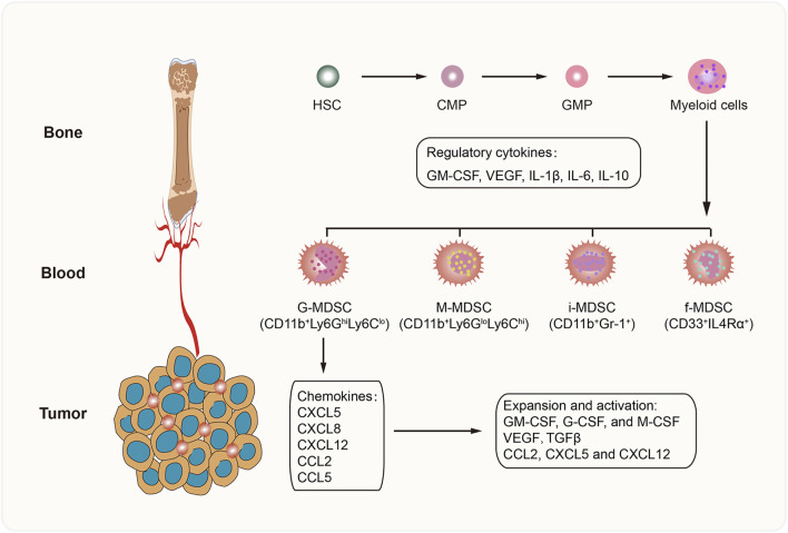
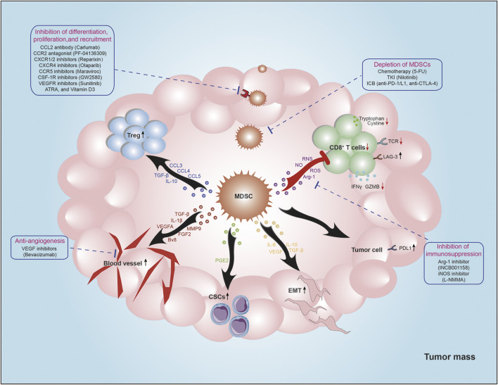
Myeloid-derived suppressor cell (MDSC) is a heterogeneous population of immature myeloid cells, has a pivotal role in negatively regulating immune response, promoting tumor progression, creating pre-metastases niche, and weakening immunotherapy efficacy. The underlying mechanisms are complex and diverse, including immunosuppressive functions (such as inhibition of cytotoxic T cells and recruitment of regulatory T cells) and non-immunological functions (mediating stemness and promoting angiogenesis). Moreover, MDSC may predict therapeutic response as a poor prognosis biomarker among multiple tumors. Accumulating evidence indicates targeting MDSC can reverse immunosuppressive tumor microenvironment, and improve therapeutic response either single or combination with immunotherapy. This review summarizes the phenotype and definite mechanisms of MDSCs in tumor progression, and provide new insights of targeting strategies regarding to their clinical applications.
Keywords: EMT; MDSC; Treg; angiogenesis; immunotherapy.
Copyright © 2021 Cheng, Yuan, Zhu and Jia.
Conflict of interest statement
The authors declare that the research was conducted in the absence of any commercial or financial relationships that could be construed as a potential conflict of interest.
Figures
References
-
- Aguilera K. Y., Rivera L. B., Hur H., Carbon J. G., Toombs J. E., Goldstein C. D., et al. (2014). Collagen Signaling Enhances Tumor Progression after Anti-VEGF Therapy in a Murine Model of Pancreatic Ductal Adenocarcinoma. Cancer Res. 74 (4), 1032–1044. 10.1158/0008-5472.can-13-2800 - DOI - PMC - PubMed
-
- Beury D. W., Parker K. H., Nyandjo M., Sinha P., Carter K. A., Ostrand-Rosenberg S. (2014). Cross-Talk Among Myeloid-Derived Suppressor Cells, Macrophages, and Tumor Cells Impacts the Inflammatory Milieu of Solid Tumors. J. Leukoc. Biol. 96 (6), 1109–1118. 10.1189/jlb.3a0414-210r - DOI - PMC - PubMed
-
- Bingisser R. M., Tilbrook P. A., Holt P. G., Kees U. R. (1998). Macrophage-Derived Nitric Oxide Regulates T Cell Activation via Reversible Disruption of the Jak3/STAT5 Signaling Pathway. J. Immunol. 160 (12), 5729–5734. - PubMed
Publication types
LinkOut - more resources
Full Text Sources