. 2021 Dec 24:8:789493.
doi: 10.3389/fcvm.2021.789493. eCollection 2021.

A Modified Surgical Ventricular Reconstruction in Post-infarction Mice Persistently Alleviates Heart Failure and Improves Cardiac Regeneration

Affiliations

A Modified Surgical Ventricular Reconstruction in Post-infarction Mice Persistently Alleviates Heart Failure and Improves Cardiac Regeneration

Siyuan Ma et al. Front Cardiovasc Med. .

Abstract

Objectives: Large ventricular aneurysm secondary to myocardial infarction (MI) results in severe heart failure (HF) and limits the effectiveness of regeneration therapy, which can be improved by surgical ventricular reconstruction (SVR). However, the conventional SVR procedures do not yield optimal long-term outcome in post-MI rodents. We hypothesized that a modified SVR procedure without aggressive purse string suture would persistently alleviate HF and improve cardiac regeneration in post-MI mice. Methods: Adult male C57 mice were subjected to MI or sham surgery. Four weeks later, mice with MI underwent SVR or 2nd open-chest operation alone. SVR was performed by plicating the aneurysm with a single diagonal linear suture from the upper left ventricle (LV) to the right side of the apex. Cardiac remodeling, heart function and myocardial regeneration were evaluated. Results: Three weeks after SVR, the scar area, LV volume, and heart weight/body weight ratio were significantly smaller, while LV ejection fraction, the maximum rising and descending rates of LV pressure, LV contractility and global myocardial strain were significantly higher in SVR group than in SVR-control group. The inhibitory effects of SVR on LV remodeling and HF persisted for at least eight-week. SVR group exhibited improved cardiac regeneration, as reflected by more Ki67-, Aurora B- and PH3-positive cardiomyocytes and a higher vessel density around the plication area of the infarcted LV. Conclusions: SVR with a single linear suture results in a significant and sustained reduction in LV volume and improvement in both LV systolic and diastolic function as well as cardiac regeneration.

Keywords: heart failure; myocardial infarction; myocardial regeneration; surgical ventricular reconstruction; ventricular aneurysm.


Conflict of interest statement

The authors declare that the research was conducted in the absence of any commercial or financial relationships that could be construed as a potential conflict of interest.

Figures

Figure 1
Figure 1
Mouse MI model characterized by serious heart failure with enlarged left ventricle (LV) and aneurysm. (A) Representative electrocardiogram showing classical ST-segment elevation in Lead II in response to left coronary artery ligation. (B) Infarct area (IA) determined by triphenyltetrazolium chloride (TTC) staining at 6 h after MI. Red staining area indicates survival myocardium, pale area indicates infarct area. Scale bar = 5 mm. (C) Mortality in the 1st week after MI and the survived MI mice after 1 week could survive to 4 weeks. (D) Representative image of LV aneurysm at 4 weeks after MI surgery (100% incidence). (E) Representative recordings of 2D and M-mode echocardiographic images of sham and MI mice at 4 weeks after surgery. Normal sham LV and enlarged LV of MI heart was indicated (red line). (F) LV ejection fraction (LVEF) and LV end systolic volume index (LVESVI) at 4 weeks after surgery. (G) Real-time PCR for myocardial natriuretic peptide type A (Nppa) and natriuretic peptide type B (Nppb) expression at 4 weeks after surgery. *P < 0.05 MI group vs. sham group. MI, myocardial infarction; Data are means ± SEM.
Figure 2
Figure 2
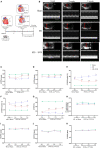
Effects of SVR surgery on remodeling and function of LV and RV evaluated by a time-course monitoring of Echocardiography from 1 to 3 weeks after SVR or 2nd chest-opening alone. (A) Schematic of MI and SVR mouse model. (B) Representative recordings of echocardiographic images of LV. The red lines indicate LV diameter. (C) LV systolic posterior wall thickness (LVPWs). (D) LV diastolic posterior wall thickness (LVPWd). (E) LV end systolic volume index (LVESVI). (F) LV end diastolic volume index (LVEDVI). (G) LV ejection fraction (LVEF). (H) The pulmonary valve (PV) peak pressure gradient. (I) RV systolic free wall thickness (RVFWs). (J) RV diastolic free wall thickness (RVFWd). For C-J, n=10 in each group. (K) Tricuspid annular plane systolic excursion (TAPSE), n = 5 in each group. *P < 0.05 between MI group and sham group; #P < 0.05 between MI+SVR group and sham group; &P < 0.05 between MI+SVR group and. MI group. +P<0.05 vs. the initial value at 4 weeks after MI. MI, myocardial infarction; SVR, surgical ventricular reconstruction; LCA, left coronary artery; LVA, left ventricular aneurysm; COA, chest-opening alone; LV, left ventricle; RV, right ventricle; Data are means ± SEM.
Figure 3
Figure 3
Effects of SVR on myocardial deformation of LV measured by speckle-tracking echocardiography at 3 weeks after SVR surgery. (A) Representative images of longitudinal and circumferential views of LV in each group. (B) Global longitudinal strain (GLS) in endocardium and epicardium. (C) Global radial strain (GRS) at longitudinal and circumferential view, respectively. (D) Global circumferential strain (GCS) in endocardium and epicardium. (E) Fractional area change (FAC). For B-E, n = 10 in each group. *P < 0.05 vs. sham 7w group; #P < 0.05 vs. sham 7w group; &P < 0.05 vs. MI 7w group. Data are means ± SEM. All the strain values were negative, indicating the direction of myocardial motion. To facilitate comparison, we used the absolute values in each group to plot. MI, myocardial infarction; SVR, surgical ventricular reconstruction; LV, left ventricle.
Figure 4
Figure 4
Effects of SVR on systolic and diastolic function of LV evaluated by invasive LV hemodynamics at 3 weeks after SVR surgery. (A) Representative pressure curve recordings of LV pressure and the rate of change of LV pressure. (B) LV systolic pressure (LVSP) and end-diastolic pressure (LVEDP). (C) Maximum rising rate of the LV pressure (dp/dt max) and maximum descending rate of the LV pressure (dp/dt min). (D) LV contractility. (E) Exponential time constant of relaxation (τ). For B-E, n = 5 in each group. *P < 0.05 vs. sham 7w group; #P < 0.05 vs. sham 7w group; &P < 0.05 vs. MI 7w group. Data are means ± SEM. MI, myocardial infarction; SVR, surgical ventricular reconstruction; LV, left ventricle.
Figure 5
Figure 5
Effects of SVR on cardiac remodeling evaluated by histological analysis and biomarkers of heart failure at 3 weeks after SVR surgery. (A) Representative pictures of the whole heart in each group. Larger heart size and LV aneurysm are visible in MI 7w group rather than in MI 7W + SVR 3W group. (B) The heart weight to body weight ratio (HW/BW). (C) The heart weight to tibia length ratio (HW/TL). (D) Azan-Masson staining of the heart at longitudinal section in MI 7w and MI 7w + SVR 3w groups. The old infarct size (IS) was calculated by scar (blue staining) size/LV circumference size. (E) Real-time PCR for myocardial Nppa and Nppb gene expression. *P < 0.05 vs. sham 7w group; #P < 0.05 vs. sham 7w group; &P < 0.05 vs. MI 7w group. Data are means ± SEM. MI, myocardial infarction; SVR, surgical ventricular reconstruction; LV, left ventricle; Nppa, natriuretic peptide type A; Nppb, natriuretic peptide type B.
Figure 6
Figure 6
Effects of SVR on cardiomyocyte proliferation at the border or plication area at 3 weeks after 2nd surgery. (A) Ki67 positive-staining cardiomyocytes (CMs). (B) Aurora B positive-staining CMs. (C) pH3 positive-staining CMs. Bar = 25 um. &P < 0.05 vs. MI 7w group. Data are means ± SEM. MI, myocardial infarction; SVR, surgical ventricular reconstruction. In each of the two side-by-side figures, the right figure is an enlarged view of the white box in the left figure.
Figure 7
Figure 7
Effects of SVR on angiogenesis at the border or plication area at 3 weeks after 2nd surgery. (A) Representative pictures showing α-SMA positive staining vessels. (B) Semi-quantitation of α-SMA positive staining vessels. (C) Expression levels of vascular endothelial growth factor alpha (VEGFα) and platelet-derived growth factor (PDGF). Data are means ± SEM. &P < 0.05 vs. MI 7w group, #P < 0.05 vs. sham group. MI, myocardial infarction; SVR, surgical ventricular reconstruction. α-SMA, smooth muscle actin. In each of the two side-by-side figures, the right figure is an enlarged view of the white box in the left figure.

Similar articles

Cited by

References

    1. Bahit MC, Kochar A, Granger CB. Post-myocardial infarction heart failure. JACC Heart Fail. (2018) 6:179–86. 10.1016/j.jchf.2017.09.015 - DOI - PubMed
    1. Bolognese L, Neskovic AN, Parodi G, Cerisano G, Buonamici P, Santoro GM, et al. . Left ventricular remodeling after primary coronary angioplasty: patterns of left ventricular dilation and long-term prognostic implications. Circulation. (2002) 106:2351–7. 10.1161/01.CIR.0000036014.90197.FA - DOI - PubMed
    1. Ruzza A, Czer LSC, Arabia F, Vespignani R, Esmailian F, Cheng W, et al. . Left ventricular reconstruction for postinfarction left ventricular aneurysm: review of surgical techniques. Tex Heart Inst J. (2017) 44:326–35. 10.14503/THIJ-16-6068 - DOI - PMC - PubMed
    1. Di Donato M, Castelvecchio S, Menicanti L. End-systolic volume following surgical ventricular reconstruction impacts survival in patients with ischaemic dilated cardiomyopathy. Eur J Heart Fail. (2010) 12:375–81. 10.1093/eurjhf/hfq020 - DOI - PubMed
    1. Athanasuleas CL, Buckberg GD, Stanley AW, Siler W, Dor V, Di Donato M, et al. . Surgical ventricular restoration in the treatment of congestive heart failure due to post-infarction ventricular dilation. J Am Coll Cardiol. (2004) 44:1439–45. 10.1016/j.jacc.2004.07.017 - DOI - PubMed